Download
1 / 22
260 likes | 389 Views
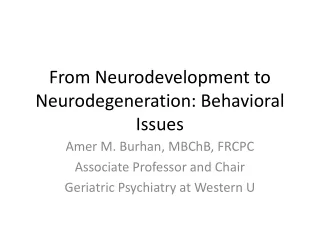
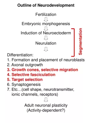
Outline of Neurodevelopment. Fertilization. Embryonic morphogenesis. Induction of Neuroectoderm. Segmentation. Neurulation. Differentiation: 1. Formation and placement of neuroblasts 2. Axonal outgrowth 3. Growth cones, selective migration 4. Selective fasciculation 5. Target selection

E N D
Outline of Neurodevelopment Fertilization Embryonic morphogenesis Induction of Neuroectoderm Segmentation Neurulation Differentiation: 1. Formation and placement of neuroblasts 2. Axonal outgrowth 3.Growth cones,selective migration 4.Selective fasciculation 5.Target selection 6. Synaptogenesis 7. Etc…(cell shape, neurotransmitter, ionic channels, receptors) Adult neuronal plasticity (Activity-dependent?)
WT Drosophila retina Soap bubbles (4 cells) (5 cells) (1 cell) (2 cells) Drosophila Rough eye (Roi) mutants (3 cells) (6 cells) (Hayashi and Carthew, 2004) Physical forces of surface contacts largely determine cell shape: Drosophila cone cell morphology modeled by soap bubbles!
Selective Adhesion Determines Specificity of Tissue and Cellular Associations
Epidermis + Mesoderm 1. Sponges (Wilson, 1907) 2. Amphibians (Townes and Holtfretter, 1955) 3. Chick (Moscona, 1952) (Townes and Holtfretter, 1955) Selective Aggregation of dissociated embryonic tissues (vertebrate and invertebrate) suggests ancient (surface) Adhesion Molecules
Experimental recreation of morphogenesis by mixing cells expressing low and high levels of one surface adhesion gene (N-cadherin) +4 hrs +24 hrs Green = high N-cadherin Red = low N-cadherin (Foty and Steinberg, 2004)
N T D V T D V N (Friche,et al. 2001) Retinotectal Mapping Visualized by Dye Injection in Zebrafish
A (T) P (N) L(V) dorsal ventral M(D) nasal temporal Do Molecular Cues Determine the Retinotectal Spatial-topic Map? Optic tectum A (T) D T N M (D) L (V) V P (N) Retina Optic Tectum
Subjective “down” Subjective “up” Rotate Eye 180o Retina Optic Tectum (T) V Subjective “down” N T (V) (D) D (N) Retinotectal Map is Preserved Despite Experimental Rotation of the Eye: “Chemaffinity Hypothesis” (Sperry, 1956) (T) D T N (V) (D) V (N)
Longitudinal Tracts MP1 aCC Q1 pCC MP1 Q1 MP1 aCC Q1 pCC Identified Neurons Commissural Tracts Grasshopper embryo Q1 (Meyers and Bastiani, 1993) Early Embryonic Insect Neurons form a Repeated Segmental Scaffold: Favorable preparation for studying axonal guidance
Pioneer Neurons Create the Early Scaffold of the Adult Nervous System growth cone pioneer neuron guidepost cells selective fasciculation Pioneer neuron and guidepost cells may die after pathway is pioneered, by apoptosis
Guidepost Cells Growth Cone Pioneer Neurons Pioneer Neurons and Guidepost Cells guide the initial path of peripheral nerve tracts in embryonic grasshopper limbs CT1 Photoablated Control (Bentley and Caudy, 1983)
filapodia F-actin Tubulin lamellipodia 2nd Messengers Extracellular Cues Ca+2 Cytoskeletal Rearrangment Intracellular Signaling Pathways GTP cAMP Growth Cones are DynamicSensory Organelles that Guide the Growth of Embryonic Axons (Forscher lab) • Sensing and Transducing: • Diffusible Cues • Contact-dependent Cues • Trophic Factors • Neurotransmitters (Play GFP-Actin Growth Cone Movie) Dr. Andrew Matus Friedrich Miescher Institute, Switzerland
Identification of Molecules Mediating Axonal Guidance using Model Systems 1. Biochemical approach: Friedrich Bonhoeffer, retinotectal culture assay. Observe Neuronal Specificity Functional Assay Fractionate Native Factors Temporal Nasal Purify and Identify Factor (Ephrins...) Nasal Axons Temporal Axons
Observe WT Neuronal Specificity Screen for Mutants of Neuronal Specificity Clone Mutant Genes Identify Factors (Semphorins, Slit, Robo, Commissureless...) Identification of Molecules Mediating Axonal Guidance using Model Systems 2. Molecular genetic approach: Corey Goodman, Drosophila screens for neurodevelopmental defects.
Conserved Structural Classes of Axonal Guidance Molecules:Modular Construction and Multifunctionality 1. Laminin, fibronectin and extracellular matrix proteins. 2. Cadherins and catenins. (Ca+2 dependent) 3. Cell adhesion molecules (CAMs) (containing IgG domains). 4. Receptor tyrosine kinases and receptor phosphatases.
(Secreted) (sema, slit) (netrin) (fas) (eph) (Membrane Associated) Functional Classes of Axonal Guidance Molecules Molecules may function for both: 1. Selective adhesion 2. Intracellular signaling
diffusible repellant diffusible attractant Contact-dependent attractant Contact-dependent repellant selective fasciculation Axonal Guidance Cues (Timing is critical)
Axonal Guidance 1. Pioneer neurons construct the earliest scaffold of the nervous system, following chemical cues. 2. Multiple chemical cues guide growth cones, including long-range diffusible cues (secreted molecules) and short-range contact mediated cues (membrane associated). 3. Chemical cues may be attractive or repulsive. 4. Chemical cues mediate both selective adhesion and intercellular signaling. 5. Axonal guidance molecules are ancient conserved molecules, including a large class with structural similarity to immunoglobulins. 6. Final axonal pathways likely specified by unique combinations of molecular cues expressed by growing neurons and targets (Sperry’s Chemoaffinity Hypothesis). 7. Human mutations of axonal guidance genes may underlie many hereditary neurological conditions affecting complex cognitive functions.
WT WT ast ast WT WT WT ast Drosophila robo disrupts longitudinal tract formation Zebrafish ROBO Mutant (astray) Disrupts Midline Retinotectal Axonal Projections (Fricke, et al. 2001) Robo acts as a receptor for a midline repulsive cue
Normal (horizontal gaze palsy) HGPPS (reduced hindbrain volume) (scoliosis) (Jen, et al., 2004) Drosophila robo disrupts longitudinal tract formation Human ROBO Mutation causes HGPPS (Horizontal Gaze Palsy with Progressive Scoliosis)
The Axon Guidance Receptor Gene ROBO1 Is a Candidate Gene for Developmental Dyslexia Katariina Hannula-Jouppi1, Nina Kaminen-Ahola1, Mikko Taipale1,2, Ranja Eklund1, Jaana Nopola Hemmi1,3, Helena Kaariainen4,5, Juha Kere1,6* 1 Department of Medical Genetics, University of Helsinki, Finland, 2 European Molecular Biology Laboratory, Gene Expression Programme, Heidelberg, Germany, 3 Department of Pediatrics, Jorvi Hospital, Espoo, Finland, 4 Department of Medical Genetics, The Family Federation of Finland, Helsinki, Finland, 5 Department of Medical Genetics, University of Turku, Turku, Finland, 6 Department of Biosciences at Novum and Clinical Research Centre, Karolinska Institutet, Stockholm, Sweden PLOS Genetics (2005) 1: 0467
Development Proceeds by Progressive Developmental Restrictions (pluripotent) (differentiated)