Download
1 / 70
710 likes | 1.01k Views
Neurons and the General Layout of the Nervous System. Ch. 3. Outline. General Layout of the Nervous System The Meninges, Ventricles, and the Blood-brain Barrier Cells of the Nervous System Neuroanatomical Techniques. General Layout of the Nervous System.

E N D
Outline • General Layout of the Nervous System • The Meninges, Ventricles, and the Blood-brain Barrier • Cells of the Nervous System • Neuroanatomical Techniques
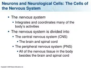
2 divisions along several different criteria • Central Nervous System vs. Peripheral Nervous System: the CNS is within the bony skull and vertebral column • brain vs. spinal cord: comprise the 2 parts of the CNS • somatic vs. autonomic: comprise the 2 parts of the PNS. The somatic branch interacts with the external environment; the autonomic branch interacts with the internal environment (regulating)
2 divisions along several different criteria • efferent vs. afferent: two branches of the somatic and autonomic. Refers to whether PNS nerves bring sensory information into the CNS (afferent) or carry motor commands away from the CNS (efferent) • sympathetic vs. parasympathetic: the two kinds of efferent nerves of the autonomic division of the PNS. Sympathetic activation arouses an organism, parasympathetic activation relaxes an organism
The Meninges • the brain and spinal cord are well-protected by the skull and vertebrae, and by three membranes called the meninges: (1) the dura mater (tough mother; outside) (2) the arachnoid membrane (spidery; middle) (3) the pia mater (gentle mother; inside)
Ventricles • cerebrospinal fluid (CSF) is manufactured by choroid plexuses, which are capillary networks that protrude into the ventricles • CSF supports and cushions the brain
Ventricles • CSF circulates through the ventricular system of the brain (“hollow” parts of the brain), the central canal of the spinal cord, and the subarachnoid space; and it is absorbed into large channels called sinuses in the dura mater and then into the blood stream
Ventricles • When the flow of CSF is blocked, hydrocephalus results • This is because the choroid plexuses (small blood vessels) are continually producing CSF, but it can’t get back to the blood stream, so there is a build up, resulting in pressure on the brain
The Blood-Brain Barrier • most blood vessels of the brain do not readily allow compounds to pass from the general body circulation into the brain; this protection, called the blood-brain barrier, is due to the tightly-packed nature of the cells of these blood vessels
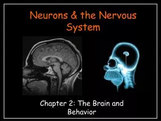
Cells of the Nervous System • the gross structures of the nervous system are made up of hundreds of billions of different cells that are either: (1) Neurons (2) Glia
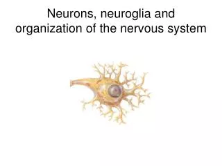
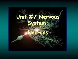
Neurons • the fundamental functional unit of the nervous system; cells that are specialized for the reception, conduction, and transmission of electrochemical signals
Neurons • most of you have seen a schematic drawing of a multipolar motor neuron; don’t be mislead by its familiar shape, as neurons come in a wide variety of sizes and shapes. The following are its nine parts: (label diagram in class)
Neurons (1) semipermeable cell membrane - (only some molecules can get through into the cell) This is because of special proteins that allows chemicals to cross the membrane; this semipermeability is critical to the normal activity of the neuron. The inside of the cell is filled with cytoplasm.
Neurons (2) cell body (soma) - the metabolic center of the cell. The soma also contains the nucleus of the neuron, which contains cell’s DNA. (3) Dendrites - shorter processes emanating from the cell body that receive information from synaptic contacts with other neurons.
Neurons (4) a single axon, that projects away from the cell body; this process may be as long as a meter! (5) axon hillock- the junction between cell body and axon; a critical structure in the conveyance of electrical signals by the neuron
Neurons (6) multiple myelin sheaths. These are formed by oligodendroglia in the CNS and Schwann cells in the PNS; they insulate the axon and assist in its conduction of electrical signals. (7) Nodes of Ranvier- the small spaces between adjacent myelin sheaths
Neurons (8) terminal buttons - the branch endings of the axon that release chemicals that allow the neuron to communicate with other cells (9) synapses - the points of communication between the neuron and other cells (neurons, muscle fibers)
Neurons • The type of neuron usually drawn in textbooks is called a multipolar neuron, because it has multiple dendrites and an axon extending from soma. There are also unipolar neurons (1 process combining both axon and dendrites off of the soma), bipolar neurons ( a single axon and a single dendrite off the soma) and interneurons that have no axons at all
Glial Cells and Satellite Cells • the most common type of cells in the nervous system are glial and satellite cells; they outnumber neurons by as much as 10:1 • glial cells are found in the CNS and satellite cells in the PNS; they provide both physical and functional support to neurons
Glial Cells and Satellite Cells • the glial cells and satellite cells that form the myelin sheaths of axons in the CNS and PNS are oligodendroglia and Schwann cells, respectively • Only Schwann cells are regenerative. Damage is permanent if it occurs in oligodendroglia (cause of Parkinson’s, degeneration of myelin of dopaminergic neurons)
Glial Cells and Satellite Cells • Researchers have begun to appreciate that glial and satellite cells play a key role in the function of the nervous systems; they help send chemical signals between neurons and they help to establish and maintain connections between neurons
Neuroanatomical Techniques • research on the anatomy of the nervous system depends upon a variety of techniques that permit a clear view of different aspects of neural structure • These techniques include:
Neuroanatomical Techniques • Golgi Stain: dye permitted individual neurons to be studied for the first time (silver chromate, only silhouette) • Nissl Stain: dye highlights cell bodies of all neurons; allowed estimation of cell density in tissue
Neuroanatomical Techniques (3) Electron Microscopy: allows visualization of the neural ultrastructure by coating with electron-absorbing substance taken up differentially by different parts of the neuron. Then pass beam of electrons through tissue onto photo paper to get image
Neuroanatomical Techniques (4) Myelin Stain: highlight myelinated pathways; less useful for studying individual axons (5) Tract Tracing: highlight individual axons; may be retrograde (trace back from terminal fields) or anterograde (trace from soma to terminal fields) after a few days, brain is sliced and treated for identifying chemical of interest (break)
The Gross Anatomyof The Nervous System Ch. 3 (cont’d)
Outline (1) Orientation and Direction in the Vertebrate Nervous System (2) The Spinal Cord (3) The Five Major Divisions of the Brain
Orientation and Direction in the Vertebrate Nervous System • First axis: anterior means toward the nose or front; posterior means towards the tail or back • Second axis: dorsal is towards the surface of the back or top of the head (as in dorsal fin); ventral indicates the surface of the chest or bottom of the head • Third axis: medial is toward the midline of the body; lateral indicates outside or away from the midline
Orientation and Direction in the Vertebrate Nervous System • Superior and inferior are often used to refer to the top and bottom of the head, respectively.
Orientation and Direction in the Vertebrate Nervous System • Planes of the brain (diagram in class): • horizontal sections • frontal (coronal) sections • sagittal sections ( a section cut between the two hemispheres is called a midsaggittal section)
The Spinal Cord • in cross section, the gray matter (cell bodies) forms a butterfly inside of the white matter (myelinated axons) • the upper (dorsal; posterior) wings of the butterfly are called dorsal horns; the lower (ventral; anterior) wings are called the ventral horns
The Spinal Cord • 31 pairs of nerves are attached to the spinal cord; as they near the cord, they split into dorsal roots (sensory axons; cell bodies lie just outside the spinal cord in the dorsal root ganglia) or ventral roots (motor axons; cell bodies lie in the ventral horns)
The Five Major Divisionsof the Brain • there are five divisions of the mammalian brain; in general higher structures are less reflexive and more complex functions, and they are more recently evolved • the nervous system is first recognizable in the developing embryo as the neural tube • the brain develops from three swellings at anterior end of the neural tube: the hind brain, the midbrain, and the forebrain
The Five Major Divisionsof the Brain • The hind brain develops into the myelencephalon and the metencephalon • The midbrain develops into the mesencephalon • The forebrain develops into the diencephalon and telencephalon (telencephalon is also called the cerebral hemispheres)