Download
1 / 34
430 likes | 1.29k Views
Pulmonary artery catheter for cardiac pressure monitoring and its role in anesthetic practice. Dr. Shalini Saini. University College of Medical Sciences & GTB Hospital, Delhi. Pulmonary artery catheter . Introduction Insertion technique Indications Complications

E N D
Pulmonary artery catheter for cardiac pressure monitoring and its role in anesthetic practice Dr. ShaliniSaini University College of Medical Sciences & GTB Hospital, Delhi
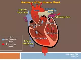
Pulmonary artery catheter Introduction Insertion technique Indications Complications Abnormal pulmonary artery and wedge pressure waveform
Introduction • In 1970, Swan,Ganz and colleague introduced pulmonary artery catheter
Pulmonary artery catheter Standard : 7-9 Fr circumference 110cm length at 10cm intervals Four internal lumen- 1. Distal-pulmonary artery pressure monitoring 2. Proximal-30cm for CVP monitoring 3. It leads to balloon near the tip 4. It houses wires for a temperature thermistor
Uses of pulmonary artery catheter • Assessment of volume status in patients undergoing major surgeries • Cardiac output measurement by thermodilution technique • Various hemodynamic parameters-pulmonary artery pressure, pulmonary capillary wedge presssure,CVP,systemic vascular resistance, pulmonary vascular resistance. • Respiratory or oxygen transport measurement-mixed venous oximetry.
Insertion Sites of insertion • Right internal jugular vein (preferred) • Left internal jugular vein (2nd choice) • Subclavian vein (disadvantages) • External jugular vein ( superficial location but tortous) • Antecubital vein • Femoral vein *After successful venous cannulation, there might be difficulty in advancement of the catheter due to abnormal venous anatomy. - most common: persistence of left superior vena cava.
Distances to right atrium, right ventricle and pulmonary artery
Wave form recorded during passage of pulmonary artery catheter Right atrial pressure resembles central venous pressure waveform Right ventricular pressure shows higher systolic pressure Pulmonary artery pressure shows diastolic step up Pulmonary artery wedge pressure similar morphology as right atrial pressure but a,c v waves appear later
Temporal relationships between systemic arterial pressure,pulmonary artery pressure, central venous pressure and pulmonary artery wedge pressure -PAP upstroke precedes radial artery pressure upstroke -Wedge pressure a wave follows ECG R wave
Three-zone model of pulmonary vasculature -described by West and colleague -tip of PAC should lie in zone3 -supine position favours zone3 condition
Indications • Patients undergoing cardiac surgery with • Poor left ventricular compliance(ejection fration <0.4, LVEDP>18mm hg) • Left wall motion abnormality • Recent MI (<6 Months) • Left main coronary lesion • Valvular lesion • Presence of pulmonary artery hypertension
2. Major procedures involving large fluid shifts or blood loss in patients with- • Cardiogenic or septic shock or with multiple organ failure • Hemodynamic instability requiring ionotropes or intra-aortic balloon counterpulsation • Hepatic transplantation • Massive ascites requiring major surgery • Surgery of aorta requiring cross-clamping • Large abdomino-perineal resection etc.
3. Intensive care unit • To measure pulmonary artery and pulmonary capillary wedge pressure • To measure cardiac output by thermodilution • To obtain intracavitary electrocardiogram • To perform atrial or ventricular pacing • To allow infusion of drugs • To perform angiography • To detect venous air embolism 4. Continous mixed venous oximetry - To assess the adequacy of perfusion
Recommendations for perioperative use of PACs(AHA 2007 guidelines) • Class 2b-(level of evidence:B) Use of PAC is reasonable in patients at risk for major hemodynamic disturbances easily detected by PAC. However, decision must be based on three parameters- a. disease b. surgical procedure c. practice setting (Experience & interpretation) • Class 3-(level of evidence:A) Routine use of PAC perioperatively, especially low risk of hemodynamic disturbances, is not recommended.
Pulmonary Artery Catheterisation and outcome controversies • PAC use in 5735 patients in first 24 hrs intensive care associated with increased mortality, hospital stay and cost.(Connors etal,1997) • Three trials including 3468 patients showed no effect on mortality but higher incidence of adverse effects(Harvey etal,2005; The ESCAPE Trial,2005; Sandham etal,2003) • A review of 53312 patients from National Trauma Data Bank showed -No mortality benefit in patients treated with PAC -Injury scale greater: mortality decreased(Friese etal,2006) • In mixed medical and surgical population,APACHE scores <25 -increased mortality >31 -significant benefit (Chittock etal,2004)
A group of experienced cardiac anesthesiologists and surgeons blinded to information from pulmonary artery catheterisation during CABG surgery were unaware of 65% of severe hemodynamic abnormalities.(Waller and Kaplan) • ICU physicians were unable to accurately predict hemodynamic data on clinical grounds and 60% made at least one change in therapy and 33% changed their diagnosis based on PAC data.(Iberti and Fisher)
Contraindications (Kaplan) Absolute contraindications • Tricuspid or pulmonary stenosis • Right atrial or ventricular mass • Tetralogy of Fallot Relative contraindications • Severe arrhythmias • Coagulopathy • Newly inserted pacemaker wires
Complications • Catheterisation a. Arrythmia-primary complication -most common premature ventricular contractions -ventricular fibrillation b. Right bundle branch block c. Complete heart block Treatment- balloon deflated and catheter withdrawn to right atrium
Catheter residence a. Catheter knotting -suspected when difficulty in withdrawing -diagnosed with chest x-ray -untied by radiologist b.Thromboembolism –rising incidence -increased with antifibrinolytic drugs c. Pulmonary infarction d. Infection ,endocarditis e. Endocardial damage, cardiac valve injury f. Thrombocytopenia (heparin induced)
g. Pulmonary artery rupture -Incidence: 0.064%-0.20% -Increased risk: female sex, hypothermia, anticoagulation, advanced age, pulmonary hypertension, mitral stenosis, coagulopathy, distal placement catheter, balloon hyperinflation -Hallmark:hemoptysis and exsanguination -Treatment: 1. Resuscitation-adequate oxygenation and ventilation 2. If hemorrhage minimal,withcoagulopathy-correct coagulopathy 3. Protection of uninvolved lung by-tilting patient to affected side, placement of double lumen endotracheal tube 4. Stop hemorrhage- apply PEEP, Bronchial blockers, resection. 5. Severe hemorrhage with recurrent bleeding-transcatheter coil embolisation.
Abnormal pressure waveforms Artifact Overwedging occurs when balloon overinflated/distal migration of cathter/eccentric balloon inflation.
B.Pathophysiologic changes 1.Mitral regurgitation -Tall v waves in waveform with bifid appearance -PAP upstroke steeper -Wedge pressure prominent v wave with gradual upstroke -Wedge pressure overestimates left ventricular filling pressure -Tall v wave:hypervolemia,CHF, VSD.
2.Mitral stenosis -Mean wedge pressure increased -y descent slurred due to obstruction to bloood flow -Similar abnormalities-left atrialmyxoma, left ventricle infarction, pericardial constriction,aorticstenosis, systemic hypertension
3.Pericardial constriction -prominent a and v waves -steep x and y descent -M or W configuration in CVP trace -diastolic ‘dip and plateau’ pattern or square root sign due to early diastolic ventricular filling -mid diastolic h wave (plateau) due to interruption in flow due to restrictive shell
4.Myocardial ischemia First time used in patient with acute MI. PAP normal relatively PAWP slightly elevated PAWP morphology abnormal Tall a waves due to diastolic dysfunction
Additional uses of pulmonary artery catheter • Thermodilution cardiac output monitoring • Principle: Stewart-Hamilton equation Q = V(Tb-Ti)K1.K2 Tb(t)dt Q = Cardiac output V = Injectate volume Tb = Blood temperature Ti = Injectate temperature K1 = Density factor: (sp heat)(sp gravity)injectate (sp heat)(sp gravity)blood K2 = A computation constant which includes heat change in transit,dead space of the catheter,injection rate, adjusts units to l/min Tb(t)dt = change in blood temperature as a function of time
2. Mixed venous oximetry Svo2 = Sao2- Vo2/ Q×1.36×Hb Svo2 = mixed venous hemoglobin saturation(%) Sao2 = arterial hemoglobin saturation(%) Vo2 = oxygen consumption(ml/min) Q = Cardiac output (l/min) Hb = hemoglobin concentration(g/dl) * Mixed venous hemoglobin saturation determined by sampling from PAC either intermittently or continous
3. Right Ventricular Ejection Fraction RVEDV = SV/RVEF RVEDV = Right ventricular end diastolic volume (ml) SV = Stroke volume RVEF = Right ventricular ejection fraction 4. Derived Hemodynamic Variables SVR = MAP-CVP × 80 CO PVR = MPAP-PAWP×80 CO SVR = systemic vascular resistance PVR = pulmonary vascular resistance MAP = mean arterial pressure CVP = central venous pressure MPAP = mean pulmonary artery pressure PAWP = pulmonary artery wedge pressure CO = cardiac output
References • Ronald.D.Miller: Pulmonary artery catheter monitoring. Cardiovascular monitoring 7th ed:1297-1314. • Kaplan: Anesthesia techniques for cardiac surgical procedures;399-408. • Circulation. 2007;116:e418-e500 • Blitt: Monitoring. Pulmonary artery cathterisation;221-263.