Download
1 / 29
290 likes | 297 Views
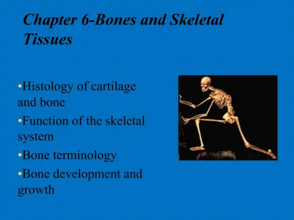
PART 2. Bones and Skeletal Tissues. Chemical Composition of Bone. 35% organic components Composed of cells, fibers, and organic substances Collagen – abundant 65% inorganic mineral salts Primarily calcium phosphate Resists compression. Bone Development.

E N D
PART 2 Bones andSkeletal Tissues
Chemical Composition of Bone • 35% organic components • Composed of cells, fibers, and organic substances • Collagen – abundant • 65% inorganic mineral salts • Primarily calcium phosphate • Resists compression
Bone Development • Ossification (osteogenesis) – bone-tissue formation • Membrane bones – formed directly from mesenchyme • Intramembranous ossification • Other bones – develop initially from hyaline cartilage • Endochondral ossification
Intramembranous Ossification Figure 6.9, steps 1–2
Intramembranous Ossification Figure 6.9, steps 3–4
Endochondral Ossification • All bones except some bones of the skull and clavicles • Bones are modeled in hyaline cartilage • Begins forming late in the second month of embryonic development • Continues forming until early adulthood
Stages in Endochondral Ossification Figure 6.10
Anatomy of Epiphyseal Growth Areas • In epiphyseal plates of growing bones • Cartilage is organized for quick, efficient growth • Cartilage cells form tall stacks • Chondroblasts at the top of stacks divide quickly • Pushes the epiphysis away from the diaphysis • Lengthens entire long bone
Anatomy of Epiphyseal Growth Areas • Older chondrocytes signal surrounding matrix to calcify • Older chondrocytes then die and disintegrate • Leaves long trabeculae (spicules) of calcified cartilage on diaphysis side • Trabeculae are partly eroded by osteoclasts • Osteoblasts then cover trabeculae with bone tissue • Trabeculae finally eaten away from their tips by osteoclasts
Organization of Cartilage within Epiphyseal Plate of Growing Long Bone Figure 6.11
Postnatal Growth of Endochondral Bones • During childhood and adolescence • Bones lengthen entirely by growth of the epiphyseal plates • Cartilage is replaced with bone CT as quickly as it grows • Epiphyseal plate maintains constant thickness • Whole bone lengthens
Hormonal Regulation of Bone Growth • Growth hormone – produced by the pituitary gland • Stimulates epiphyseal plates • Thyroid hormone – ensures that the skeleton retains proper proportions • Sex hormones (estrogen and testosterone) • Promote bone growth • Later induces closure of epiphyseal plates
Postnatal Growth of Endochondral Bones • As adolescence draws to an end • Chondroblasts divide less often • Epiphyseal plates become thinner • Cartilage stops growing • Replaced by bone tissue • Long bones stop lengthening when diaphysis and epiphysis fuse
Bone Remodeling • Bone is dynamic living tissue • 500 mg of calcium may enter or leave the adult skeleton each day • Cancellous bone of the skeleton is replaced every3 – 4 years • Compact bone is replaced every 10 years
Postnatal Growth of Endochondral Bones • Growing bones widen as they lengthen • Osteoblasts – add bone tissue to the external surface of the diaphysis • Osteoclasts – remove bone from the internal surface of the diaphysis • Appositional growth – growth of a bone by addition of bone tissue to its surface
Bone Remodeling • Bone deposit and removal • Occurs at periosteal and endosteal surfaces • Bone remodeling • Bone deposition – accomplished by osteoblasts • Bone reabsorption – accomplished by osteoclasts
Remodeling, Spongy Bone Figure 6.12
Osteoclast – A Bone-Degrading Cell • A giant cell with many nuclei • Crawls along bone surfaces • Breaks down bone tissue • Secretes concentrated HCl • Lysosomal enzymes are released Figure 6.13a
Repair of Bone Fractures • Simple and compound fractures • Treatment by reduction • Closed reduction • Open reduction
Stages of Healing a Fracture Figure 6.14
Common Types of Fractures Table 6.2 (1 of 3)
Common Types of Fractures Table 6.2 (2 of 3)
Common Types of Fractures Table 6.2 (3 of 3)
Disorders of Bones • Osteoporosis • Characterized by low bone mass • Bone reabsorption outpaces bone deposition • Occurs most often in women after menopause
Osteoporosis Figure 6.15
Disorders of Bones • Osteomalacia • Occurs in adults – bones are inadequately mineralized • Rickets • Occurs in children – analogous to osteomalacia
Disorders of Bones • Paget’s disease • Characterized by excessive rate of bone deposition • Osteosarcoma • A form of bone cancer
The Skeleton Throughout Life • Cartilage grows quickly in youth • Skeleton shows fewer chondrocytes in the elderly • Bones are a timetable • Mesoderm • Gives rise to embryonic mesenchyme cells • Mesenchyme • Produces membranes and cartilage • Membranes and cartilage ossify
The Skeleton Throughout Life • Skeleton grows until the age of 18–21 years • In children and adolescents • Bone formation exceeds rate of bone reabsorption • In young adults • Bone formation and bone reabsorption are in balance • In old age reabsorption predominates • Bone mass declines with age