Download
1 / 42
420 likes | 427 Views
OPTIMIZING DIAGNOSTIC NUCLEAR MEDICINE SERVICES AT BMC. Presenter: Dr Beda Likonda Chairperson: Mr. Alex Mpugi. Nuclear medicine (NM).

E N D
OPTIMIZING DIAGNOSTIC NUCLEAR MEDICINE SERVICES AT BMC Presenter: Dr Beda Likonda Chairperson: Mr. Alex Mpugi
Nuclear medicine (NM) • Nuclear medicine is a branch of Medicine that uses radioactive tracers in the form of unsealed sources for diagnosis, therapy (intervention) and laboratory testing of human diseases.
Atoms of elements • Nucleus contains protons and neutrons (Z+N) • Surrounded by a cloud of electrons orbiting • Sum of nuclear sub atomic particles makes atomic mass (A)
Stable and unstable nuclei • Stable state nuclei has optimal ratio of neutrons to protons approximately 1:1 • Unstable has either too many neutrons, or too many protons or excess energy (appropriate nucleons ratio)
Isotopes and radionuclei • Isotopes are Atoms of of a particular element existing with different numbers of neutrons (differ in atomic mass keeping same number of protons) • Not all isotopes are unstable (some doesn’t decay) • Unstable isotopes undergoes transformation to become stable by gamma, beta, alpha or positron emissions and the are radionuclei. • Radionucleiare important resources in nuclear medicine for diagnostic, intervention or laboratory studies.
Radioactivity • Alpha emissions • Beta emissions • Positrons emissions • Gamma emissions
Radioactive emissions Alpha Beta When a neutron is converted to proton Finite and limited range in tissues Higher energy beta-(P-32,Y-90), several mm range, homogeneous, large targets e.g. lymphoma nodule Useful for therapeutics Stopped by Plastic, glass, Aluminium • Highly energetic (He) particles • By very large unstable nucleus • High levels of cell killing close to deposition • Attractive for therapeutic application since has high LET,RBE and less dependent on oxygen • At-111, Bi-213 • Stopped by paper, skin, clothes
Radioactive emissions Positrons Gamma Electromagnetic radiations of extremely high frequency Ionizing radiations Useful for diagnostic studies ( SCINT/SPECT) Most common radionuclide is Technitium-99m Stopped by dense metal, concrete, Earth • Positive beta • A proton is converted to neutron • The short-lived positron emitting isotopes 11C, 13N, 15O and 18F • used for positron emission tomography
Quantity of radioactivity • Becquerel (Bq) one disintegration per second (dps) • Curies (Ci) based on the amount of radioactivity in 1 g of radium isotope 226Ra. 1 Ci = 3.7 × 10¹⁰ Bq. • Typical doses for imaging are often 37MBq which is 1mCi.
Quantity of absorbed radiations • Gray(Gy) energy ( a Joule) absorbed by one kilogram of tissue. • Rads is former SI unit, 1 Gray is 100Rads • Roentgen equivalent man (Rem) a measure of biological effect of an emission. Takes into account absorbed dose and quality factor. • Sieverts (Sv) one Sv is 100 Rem
Radiopharmaceuticals (tracers) • Are composed of a radionuclide and chemically chelated to a compound or and antibody. • A radionuclide maintains biological properties of non-radioactive atoms. • The chemical compound or the antibody has a selective physiological property to an organ-system. • Soluble can be injected, ingested or inhaled
Radiopharmaceuticals (tracers) • Non-chelated 131-I, 89-Sr(salts) • Chemical chelation MDP Technitium-99m, EDTMP-samarium-153 • Antibody/peptide (131-I tositumomab, 177-lutitium-DOTA )
Ideal radiopharmaceuticals (tracers) • Stable link with parent drug/metabolite which controls bio-distribution • Excreted rapidly through known simple routes • Should not undergo complex metabolism, excretion and dosimetry will be complex. • Similar physical half life radionuclide/drug for effective half life to be representative.
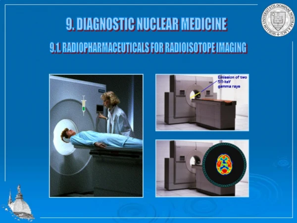
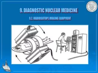
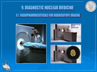
NM imaging principles • A radiopharmaceutical (tracer) is given to the patient orally, by injection, or by inhalation. • Once the tracer has distributed itself in the body, a patient is passed through a gamma camera. • Produces emission images (as opposed to transmission images), because the radioisotopes emit their energy from inside the patient. • This is a form of functional imaging as opposed to anatomical imaging by x-rays, CTscan.
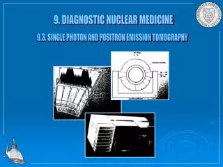
NM imaging principles • Emissions are used to create • 2-dimensions images called scintigraphy (SCINT). • 3-dimensions images called single photon emission computerized tomography (SPECT). • 3-dimensions images called positron emission tomography (PET) • 3-dimensions hybrid images, SPECT/CT ,PET/CT, SPECT/MRI or PET/MRI.
Single Photon Emission Computed Tomography “SPECT” • In SPECT, a nuclear camera records gamma-ray emissions from the patient from a series of different angles around the patient. • These projection data are used to reconstruct a series of tomographic emission images • SPECT allows physicians to better understand the precise distriburion of the radioactive agent
Positron Emission Tomography (PET) • Although more expensive than SPECT, PET has clinical advantages in certain diagnostic areas. The PET detector system is more sensitive to the PET • PET has higher spatial resolution than SPECT
Hybrid scanning techniques • The nuclear medicine scans can be superimposed, using software or hybrid cameras, on images from modalities such as CT or MRI to highlight the part of the body in which the radiopharmaceutical is concentrated. • This practice is often referred to as image fusion or co-registration, for example SPECT/CT and PET/CT. • Provides information about the anatomy and function.
Advantages of NM imaging • Ability to image the whole body in a single sitting at a comparably short time hence increased patient through fare. • Minimal radiation burden to patients, personnel and environment- the dosages used are very small • Non-invasive • Able to give information on cellular activity at an early stage
Disadvantages • Uses very costly and sophisticated equipment. • Requires highly trained personnel. • Higher chances of radiation exposure since the source is unsealed. • Radiation accidents are more likely to occur if safety precautions are compromised
Radiation protection and safety • Safe handling of radioactive materials using the principle of ALARP ( as low as reasonably practicable). This is vital in preventing unnecessary radiation from internal body organ. • Reduction of radiation doses from external sources e.g. radioactive emissions from patients, syringes, vials and waste disposal area by using the TDS rule.
TDS rule • TIME = decrease time of exposure-work as rapidly as possible in a radiation area with consistent good technique. • DISTANCE = increase distance from source about 2m from patients in patient study area during operation of instrumentation. Separate waiting areas for the injected patients and non-injected patient, relatives. • SHIELDING = lead shielding whenever possible.
Disposal of radioactive waste • Dilute and disperse in water sinks small/low dose quantities. • Store and wait for decay if high dose for at least 10 half-lives before. Separate storage for sharps vs. non-sharps. Disposal to incinerator for sharps while dumpsite for non-sharps.
At BMC • Started 2011 • Diagnostic part • Radionuclide –Technetium 99m • Pharmaceuticals • Gamma camera with ability of Scintigraphy and SPECT • Half life of technetium 6hrs • Gamma rays energy of Tc-99m is 140keV
At BMC • Dar to delivery BMC -2weeks • Number of days for effective diagnostic tests 2 weeks • 20GBq • 15GBq at BMC • Cost of procuring Mo-generator and kits 2000 Euros • List of examinations and indications
Diagnostic procedures done BMC 1.Whole body radionuclide bone scan -To detect skeletal metastases from cancers such as breast, lung, colon, thyroid, prostate. -To detect the skeleton as the primary source of cancer. -To detect inflammatory conditions of the connective tissue including infections.
Cont.... -To detect degenerative joint diseases such as osteoarthritis. -To detect sport injuries such as stress fractures -To determine whether pain from a joint prosthesis is due to loosening or infection. -To detect areas of ectopic calcification
Cont… 2.Radionuclide thyroid scan • To determine the anatomical position of the thyroid gland -To determine the functional status of the thyroid gland i.e. hypo or hyper thyroidism -To determine possibility of cancer of the thyroid gland (“cold’’ areas) - To determine outcome of radioiodine therapy
Cont… 3.Renal scans -Renogram : Will delineate the function of each kidney and the associated collecting system to provide the so-called split kidney function -Renal perfusion studies :eg after kidney transplant -Renal function :glomerular filtration rate (GFR) -Renal anatomy :e.g. ectopic kidneys
Cont… 4.Myocardial perfusion imaging :(MPI) -For diagnosis of coronary artery disease (CAD) -To differentiate between viable and non-viable myocardial tissue e.g. after a myocardial infarction (MI) hence deciding on mode of therapy (surgery vs. medical intervention) -surveillance for those patients known to be at risk of developing CAD eg diabetes
Challenges • Delays in delivery of radiopharmaceuticals e.g 99m-Tc generators from Turkey • Lack of adequate awareness of the existing NM facilities by our colleagues and the public. • Inadequate numbers of trained personnel and equipment • Lack of PET device and PET/CT hybrid devices for image fusion due to the costs involved • Global shortage of Molybdenum -99.
Optimizing services • Regular procurement of the radiopharmaceutical • Physicians should learn more about indications of the tests and benefits • Physicians produce regular requisition • Training of personnel's