Download

1 / 23
300 likes | 1.09k Views
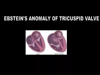
Ebstein’s anomaly. Stephanie Merhar January 21, 2011. Anatomy. Pathophysiology. Associated defects. Commonly associated with: ASD or PFO (90%) VSD, AV canal defect Pulmonary stenosis/atresia (20-25%) Wolff-Parkinson-White Syndromes: Down, Marfan, Noonan, Cornelia de Lange

E N D
Ebstein’s anomaly Stephanie Merhar January 21, 2011
Associated defects • Commonly associated with: • ASD or PFO (90%) • VSD, AV canal defect • Pulmonary stenosis/atresia (20-25%) • Wolff-Parkinson-White • Syndromes: • Down, Marfan, Noonan, Cornelia de Lange • Maternal lithium ingestion?
Epidemiology • Ebstein’s anomaly occurs in 0.3-0.8% of all congenital heart diseases • 1 in 20,000-50,000 live births • Equal male:female occurrence • Mortality in children presenting in the neonatal period is 30-50% • Mortality at all ages is 12.5%
Prenatal presentation • Difficult to diagnose prenatally • Fetal presentation is variable: possible features include cardiomegaly, RA enlargement, tricuspid regurgitation or dysplasia, arrhythmia, or fetal hydrops • Prognosis for the fetus diagnosed in utero with significant tricuspid valve disease is very poor (20% survival) • Progressive right heart dilatation • Cardiac failure • Lung hypoplasia • Pulmonary stenosis/atresia
Neonatal presentation • Congestive heart failure • Due to TR and RV dysfunction • Cyanosis • Decreased pulmonary blood flow due to R L shunt through ASD or PFO • Increased pulmonary vascular resistance in the neonatal period compounds this problem • Murmur
Physical exam • Heart sounds • First heart sound widely split with loud tricuspid component • Second heart sound usually is normal but may be widely split due to RBBB • Third and fourth heart sounds commonly present • Murmurs • Holosystolic murmur of tricuspid regurgitation
Later presentation • Cyanosis • Due to R L shunt at atrial level • Fatigue and dyspnea • Secondary to RV failure and decreased LV ejection fraction • Palpitations and sudden cardiac death • Incidental murmur • Paradoxic embolism
Arrhythmias • Due to right atrial enlargement and high prevalence of accessory pathways • 30-50% have evidence of WPW secondary to the atrialized RV tissue • Mapping and ablation are difficult • Atrial dilation disrupts anatomic landmarks • Accessory pathways are often multiple
Initial management • Prostaglandin infusion? (see next slide) • Placement of umbilical catheters • Initiation of mechanical ventilation • Minimum possible mean airway pressure • Tidal volumes of 10-15 ml/kg to overcome atelectasis • Management of pulmonary hypertension
Management of pulmonary hypertension • Nitric oxide • Reduces afterload of right ventricle • Helps distinguish functional from actual pulmonary atresia • Sedation • Other pulmonary vasodilators?
PGEs – good or bad? • Definitely need to start PGEs if functional pulmonary atresia • Need some way to get blood to lungs if going through PA is not an option • If patient gets worse on PGEs, discuss with cardiologist!
Usual postnatal evaluation • Define anatomy with echocardiography • Nature of the RV outflow tract • If pulmonary atresia/severe stenosis, likelihood of biventricular repair is very low • Great Ormond Street Ebstein (GOSE) score for severity • Ratio of combined areas of true RA plus atrialized RV to the combined areas of the functional RV, LA, and LV
Transport issues • Things to tell receiving cardiologist: • location of pulse ox (ideally pre and post), arterial blood gas, 4 point blood pressures, appearance of the CXR • Main problem to anticipate on transport • Desaturation! • Manage these babies like other babies with pulmonary hypertension
References • Aggarwal S, Chintala K, and R Humes. Sildenafil use in a symptomatic neonate with severe Ebstein’s anomaly. Am J Perinatol 2008; 25(2): 125-128. • Brown ML and JA Dearani. Ebstein malformation of the tricuspid valve: current concepts in management and outcomes. Curr Treat Options in CV Med 2009; 11:396-402. • Cherry C, Debord S, and N Moustapha-Nadler. Ebstein’s anomaly: a complex congenital heart defect. AORN Journal 2009; 89:1098-1111. • Dearani JA, O’Leary PW, and GK Danielson. Surgical treatment of Ebstein’s malformation: state of the art in 2006. Cardiol Youn 2006; 16(12-20). • Jaquiss RDB and M Imamura. Management of Ebstein’s anomaly and pure tricuspid insufficiency in the neonate. Semin Thorac Cardiovas Surg 2007;19:258-263. • Knott-Craig CJ and SP Goldbert. Management of neonatal Ebstein’s anomaly . Semin Thorac CV Surg 2007; 10:112-116. • Paranon S and P Acar. Ebstein’s anomaly of the tricuspid valve: from fetus to adult. Heart 2008; 94:237-243. • Pashia SE. Ebstein’s anomaly. Neonatal Network 2007; 26:197-207.