Download
1 / 39
440 likes | 1.56k Views
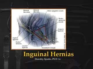
DIAPHRAGMATIC HERNIAS Maureen Austin Kimberly Novak. Diaphragmatic Hiatal Umbilical. Inguinal Femoral Perineal. Types of Hernias. Diaphragmatic Hernia. A rent in the diaphragm that allows herniation of of abdominal viscera into the thoracic cavity.

E N D
Diaphragmatic Hiatal Umbilical Inguinal Femoral Perineal Types of Hernias
Diaphragmatic Hernia • A rent in the diaphragm that allows herniation of of abdominal viscera into the thoracic cavity.
Traumatic Diaphragmatic Hernia • A common outcome of blunt force trauma to the thoracic cavity • Dogs: vehicle trauma, kicked by horse/cow • Cats: high rise trauma, vehicle trauma • Liver is the most commonly herniated • ****Young males at greatest risk- another good reason to castrate!!!!
Congenital Diaphragmatic Hernia • Pleuroperitoneal Diaphragmatic Hernia • Uncommon; autosomal recessive trait • Incomplete or failed fusion of pleuroperitonel membrane • Failure of pleuroperitoneal folds to incorporate muscle • Located dorsolateral; those on left often result in still birth or neonatal death • Stomach, spleen and small intestine most common
Congenital Diaphragmatic Hernia • Pericardioperitoneal Diaphragmatic Hernia (PPHD) • Very common –CATS • Often incidental –CATS • Not inherited, 58% males • Weimeraners predisposed (Conformation???) • Liver, omentum, spleen, falciform ligament common- NOT STOMACH
PPHD • Always congenital, most common congenital diaphragmatic hernia • Diaphragm and pericardium not continuous • Failure of septum transversum differentiation: *teratogens, genetic defect, prenatal trauma • Ventral diaphragm
Associated Abnormalities • Sternal defects/Pectus excavatum • Cranial midline abdominal wall hernia • Umbilical hernia • Cardiac defects (VSD) • PSS • Pulmonary vascular disease
Diagnostic Dilemma • 20% traumatic diagnosed @ 4 weeks post trauma:*omentum plug, rent w/no hernia, failure to dx • 48% PPHD < 1y; 36%@ 1-4y • May not be until as old as 14y: *omentum plug; formed, but weak diaphragm, • often incidental-no symptoms
Clinical Signs Asymptomatic Gravely Ill
Clinical Signs • Respiratory signs > GI signs • Dyspnea, tachypnea, coughing • Pleural effusion, pericardial effusion • Vomiting, gagging , diarrhea • Auscultation and Palpation helpful • Rare encephalopathy • Shock (both acquired and congenital) • Concurrent injuries/congenital defects
How Do You Diagnose a Diaphragmatic Hernia? • What you do first is going to depend on your history and physical examination • Is there history of trauma? • Respiratory problems? • Do you hear borborygmi on thoracic auscultation? • The tools used to diagnose a diaphragmatic hernia are the same for each type. The results obtained from your diagnostics will be different depending on the type of hernia you have.
Most often you will be using an imaging modality to confirm a diagnosis of diaphragmatic hernia • Survey radiographs • Gastrogram (upper GI series) • Positive contrast celiography
Survey Radiographs • Signs of a traumatic hernia • Loss of the diaphragmatic shadow • Presence of abdominal viscera in the thoracic cavity i.e. a radiolucent gas filled structure • Cranial and /or lateral displacement of the heart and lungs due to abdominal viscera pushing on them • Cranial and /or lateral displacement of the stomach or intestines within the abdominal cavity due to liver herniation
Survey Radiographs cont... • Signs of pericardioperitoneal hernia • Enlarged, globoid cardiac silhouette • Tubular gas shadows within the pericardium • Loss of a distinct ventral border of the cupula of the diaphragm without there being pleural fluid • A mesothelial remnant between the heart and the diaphragm • The appearance of a small liver or cranial displacement of the stomach suggesting herniation of the liver
Gastrogram (Upper GI series) • This positive contrast study will give you a definitive diagnosis of diaphragmatic hernia if the stomach or intestines are herniated into the thoracic cavity • Barium sulfate or a water soluble iodinated positive contrast medium is given by orogastic intubation and then radiographs are taken • Remember though that if you do not see any herniated organs in the thoracic cavity filled with the contrast solution it does not mean that you do not have a DH. You could either have no organs herniated through or a non-GI organ is herniated (spleen).
Positive Contrast Celiography • This procedure is usually done if survey radiographs or an upper GI study fail to give you an unequivocal diagnosis • A water soluble iodinated contrast medium is injected through a catheter into the peritoneal cavity just to the right of midline and cranial to the umbilicus. • If there is a defect in the diaphragm then the contrast material will enter into the pleural or pericardial space depending on the type of hernia • However, you can get a false negative with this test if the defect in the diaphragm is covered up by the omentum
Treatment • The definitive treatment for any of the hernias described is surgical repair of the defect in the diaphragm and replacement of any abdominal viscera that was herniated back into the thoracic cavity • Patients with traumatic hernias are first stabilized and rested before proceeding with surgery unless life-threatening hypoventilation, caused by abdominal viscera compressing the lungs, occurs
Treatment cont.. • In the pericardioperitoneal hernia repair closing of the defect in the diaphragm will also simultaneously close the opening to the pericardial sac since the two are conjoined together • Air within the thoracic cavity should be released by means of thoracentesis or a tube thoracostomy
The case of the coughing cat • Chelsea • 13 year old spayed female Burmese • Presented March 22, 2002 with a history of intermittent gagging/retching and occasional vomiting with no pattern of occurrence • Owner acquired the cat in may 1999 after the previous owners had decided to euthanize Chelsea due to erratic episodes of severe illness that could never be explained and would seem to just go away on its own • Chelsea was up to date on all vaccines and was on revolution for heartworm and flea preventative.
Physical exam findings • Temp normal • Pulse- 200beats per minute • Respiratory rate- 50 breaths per minute • weight- 7.1 lbs • Upon auscultation increased lung sounds were heard bilaterally • Abdominal palpation yielded a cough from Chelsea and tracheal palpation also caused her to begin gagging with an increase respiratory effort
Rule outs • Feline asthma • Upper Airway disease • Tracheal abnormalities • Diagnostics • Survey thoracic and abdominal radiographs • CBC and Chem profile • Fecal • Urinalysis
Fecal results • Normal Chem profile results • Creatinine 2.8 (0.7 – 2.2) • BUN 36 (18 – 41) • Total Protein 10.4 (5.5 – 7.7) CBC results • Normal Urinalysis • Normal
Survey radiographic report • Liver present in the cranial abdomen and shaped oddly • Caudal portion of the right side of thorax appears to be occupied by a well marinated gas filled structure • A stomach shadow is not seen in the normal position in the abdomen and the colon is excessively far cranial in the abdominal cavity • A dense bronchial and interstitial lung pattern is seen • Kidneys appear to be small • Recommended to do a positive contrast shallow
Gastrogram results • Results consistent with herniation of the stomach in the thoracic cavity therefore diagnosing a diaphragmatic hernia • There was no evidence of any abdominal viscera within the pericardial sac or of a connection between the diaphragm and the pericardium
What do we do now? • Chelsea was scheduled for diaphragmatic hernia repair the next morning • A perforation was found in the diaphragm located on the midline, dorsal to the liver, that measured approximately two and one half inches in length. The edges of the hernia were smooth and the tissue was healthy • The stomach and part of the spleen had herniated into the thoracic cavity and were pulled through the hole and placed back into their normal positions. Both organs looked normal • There were no adhesions and the pleura of the thorax was intact • There was no evidence of a connection between the pericardium and diaphragm
Post-surgery • Chelsea did not need any additional medications after surgery other than pain medication • Exercise was to be limited for 10 days post op to prevent her incision from becoming infected
So how did this happen to Chelsea? • There was no history of trauma • Records obtained from the previous owners showed instances where Chelsea became violently ill and then would have regression of clinical signs • She was referred to MSU prior to the exchange in ownership but no radiographs were taken at that time
Diagnosis • Chelsea was diagnosed with a congenital pleuroperitoneal diaphragmatic hernia • While this type of hernia is very rare, it most common to see the stomach, spleen, and small intestine through a left dorsolateral diaphragm defect • It was believed that this was not a traumatic hernia due to the fact that there were no adhesions within the thoracic cavity, the pleura was intact, and all organs were completely normal in appearance • Chelsea's hernia was present at birth and over the years grew in size until abdominal viscera were able to pass through. • Her intermittent gagging/vomiting was probably caused by the stomach and spleen pushing on her lungs and heart
References Biery DN., Owens JM. Radiographic Interpretation for The Small Animal Clinician 2nd Edition. Baltimore: Williams & Wilkins. 1999. Birchard SJ., Sherding RG. Saunders Manual of Small Animal Practice 2nd Edition. New York: WB Saunders & Co. 2000. Ettinger, SJ., Feldmen E.C. Textbook of Veterinary Internal Medicine 5th Edition. New York: WB Saunders & Co. 2000. Hoskins, JD. Veterinary Pediatrics: Dogs and Cats from Birth to Six Months. New York: WB Saunders & Co. 2001. Norden, DM., Lahunta, Ad. The Embryology of Domestic Animals: Developmental Mechanisms and Malformations. Williams and Wilkins 1985.