Download
1 / 27
420 likes | 1.81k Views
ABDOMINAL HERNIAS. Fadi J. Zaben RN MSN. Definition:. A hernia is a protrusion of an organ, tissue, or structure through the wall of the cavity in which it is normally contained. It is often called a rupture. The abdomen is a common place for hernias to occur .

E N D
ABDOMINAL HERNIAS Fadi J. Zaben RN MSN
Definition: • A hernia is a protrusion of an organ, tissue, or structure through the wall of the cavity in which it is normally contained. • It is often called a rupture. • The abdomen is a common place for hernias to occur. • Hernias may be present at birth due to incomplete closure of a structure, or they may develop later due to increased abdominal pressure pushing against a weakened area of muscle or its fibrous sheath (fascia).
Etiology: • Results from congenital or acquired weakness (traumatic injury, aging) of the abdominal wall. • May result from increased intra-abdominal pressure due to heavy lifting, obesity, pregnancy, straining, coughing, or proximity to tumor.
Classification: • Classification by Site. • Classification by Severity.
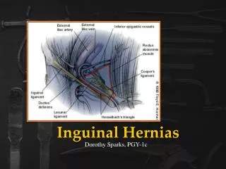
Classification by Site: • Inguinal Hernia into the inguinal canal (more common in males). • Indirect inguinal: • It occurs due to a weakness of the abdominal wall at the point through which the spermatic cord emerges in the male and the round ligament in the female. • Through this opening, the hernia extends down the inguinal canal and often into scrotum or labia. • Direct inguinal: • Itpasses through the posterior inguinal wall; more difficult to repair than indirect inguinal hernia.
Continue…….. • Femoral Hernia into the femoral canal, appearing below the inguinal ligament. • Umbilical Intestinal Protrusion at the umbilicus due to failure of umbilical orifice to close. Occurs most often in obese women, children, and in patients with increased intra-abdominal pressure from cirrhosis and ascites.
Continue….. • Ventral or Incisional Intestinal Protrusion due to weakness at the abdominal wall; may occur after impaired incisional healing due to infection or drainage. • Peristomal Hernia through the fascial defect around a stoma and into the subcutaneous tissue.
Classification by Severity: • Reducible Hernia: the protruding mass can be placed back into abdominal cavity. • Irreducible Hernia: the protruding mass cannot be moved back into the abdomen.
Continue…. • Incarcerated Hernia: an irreducible hernia in which the intestinal flow is completely obstructed. • Strangulated Hernia: an irreducible hernia in which the blood and intestinal flow are completely obstructed; develops when the loop of intestine in the sac becomes twisted or swollen and a constriction is produced at the neck of the sac.
Risk Factors for Abdominal Hernia: • Abdominal surgery. • Chronic constipation. • Chronic cough. • Enlargement of the prostate or other conditions that can lead to straining to urinate. • Family history of hernias. • Lifting or pushing heavy objects. • Male gender. • Nutritional deficiencies. • Obesity. • Overexertion. • Smoking. • Undescended testes.
Clinical Manifestations: • Bulging over herniated area appears when patient stands or strains, and disappears when supine. • Pain. • Hernia tends to increase in size and recurs with intra-abdominal pressure. • Strangulated hernia presents with pain, vomiting, swelling of hernial sac, lower abdominal signs of peritoneal irritation, fever.
Diagnostic Evaluation: Based on clinical manifestations: • Physical Examination (P/E). • Abdominal X-rays: reveal abnormally high levels of gas in the bowel. • Laboratory studies (complete blood count, electrolytes)may show hemoconcentration (increased hematocrit), dehydration (increased or decreased sodium), and elevated white blood cell (WBC) count, if incarcerated.
Management: • Mechanical (non surgical) treatment. • Surgical treatment.
Mechanical Treatment: • It is for reducible hernia only. • Truss: • It is an appliance with a pad and belt that is held snugly over a hernia to prevent abdominal contents from entering the hernial sac. • A truss provides external compression over the defect. • It should be removed at night and reapplied in the morning before patient arises. • It is used only when a patient is not a surgical candidate.
Continue….. • Peristomal hernia is often managed with a hernia support belt with Velcro, which is placed around an ostomy pouching system (similar to a truss). • Conservative measures: • No heavy lifting. • NO straining at stool. • And any measures that would increase intra-abdominal pressure should be a void.
Surgical Treatment: • It recommended to correct hernia before strangulation occurs, which then becomes an emergency situation. • Herniorrhaphy: removal of hernial sac; contents replaced into the abdomen; layers of muscle and fascia sutured. • Laparoscopic herniorrhaphyis a possibility and often performed as outpatient procedure. • Hernioplasty involves reinforcement of suturing (often with mesh) for extensive hernia repair. • Strangulated hernia requires resection of ischemic bowel in addition to repair of hernia.
Complications: • Bowel obstruction. • Recurrence of hernia.
Nursing Assessment: • Ask patient if hernia is enlarging and uncomfortable, reducible or irreducible; determine relationship to exertion and activities. • Assess bowel sounds and determine bowel pattern. • Determine if patient is exhibiting signs and symptoms of strangulation
Nursing Diagnoses: • Chronic Pain related to bulging hernia (mechanical). • Acute Pain related to surgical procedure. • Risk for Infection related to emergency procedure for strangulated or incarcerated hernia.
Nursing Interventions: Achieving Comfort: • Fit patient with truss or belt when hernia is reduced, if ordered. • Trendelenburg's position may reduce pressure on hernia, when appropriate. • Emphasize to patient to wear truss under clothing and to apply before getting out of bed when hernia is reduced. • Give stool softeners as directed. • Evaluate for signs and symptoms of hernial incarceration or strangulation. • Insert NG tube for incarcerated hernia, if ordered, to relieve intra-abdominal pressure on herniated sac.
Relieving Pain Postoperatively: • Have the patient splint the incision site with hand or pillow when coughing to lessen pain and protect site from increased intra-abdominal pressure. • Administer analgesics, as ordered. • Teach about bed rest, intermittent ice packs, and scrotal elevation as measures used to reduce scrotal edema or swelling after repair of an inguinal hernia. • Encourage ambulation as soon as permitted. • Advise patient that difficulty in urinating is common after surgery; promote elimination to avoid discomfort, and catheterize if necessary.
Preventing Infection: • Check dressing for drainage and incision for redness and swelling. • Monitor for other signs and symptoms of infection: fever, chills, malaise, diaphoresis. • Administer antibiotics, if appropriate.
Patient Education and Health Maintenance: • Advise that pain and scrotal swelling may be present for 24 to 48 hours after repair of an inguinal hernia. • Apply ice intermittently. • Elevate scrotum, and use scrotal support. • Take medication prescribed to relieve discomfort. • Teach to monitor self for signs of infection: pain, drainage from incision, temperature elevation. Also, report continued difficulty in voiding. • Inform that heavy lifting should be avoided for 4 to 6 weeks. Athletics and extremes of exertion are to be avoided for 8 to 12 weeks postoperatively, per provider instructions.