Download

1 / 60
1.28k likes | 2.87k Views
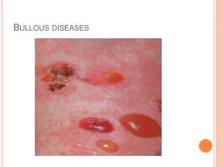
BULLOUS DISEASES. Bollous Diseases Difinitions. •Blister: collection of clear fluid. •Bulla: Blister>5mm diameter. • Vesicle: Blister<5mm diameter. • Crust Dried exudate on skin. •BSDs are skin conditions characterised by blister formation. Diagnosis of bullous Disorders.

E N D
Bollous Diseases Difinitions •Blister: collection of clear fluid. •Bulla: Blister>5mm diameter. • Vesicle: Blister<5mm diameter. • Crust Dried exudate on skin. •BSDs are skin conditions characterised by blister formation.
Diagnosis of bullous Disorders -clinical evaluation -histopathological examination -Immunoflourescent examination
Light microscopy -inflammatory infilterate -level of blister formation
Immunofluorescence -tissue-bound antibodies(direct) -circulating antibodies(indirect)
Basement membrane zone • Plasma membrane: basal keratinocyte hemidesmosome • Lamina Lucida • Lamina Densa – Type IV collagen • Anchoring fibrils – Type VII collagen
PATHOPHYSIOLOGY .The keratinocytes of the epidermis are tightly bound together by desmosomes . .Beneath the epidermis lies the BMZ, which is a specialised area of cell- extracellular matrix adhesion. .Specialised structures traversing this zone anchor the epidermis to the dermis. .These structures(matrix) comprises polysaccharides and proteins(including collagens) linked to form macromolecules(adhesion complex).
If any of these specialised attachments are malformed, absent or damaged, seperation may occur leading to accumulation of fluid in the extracellular space and blister formation. • The BMZ is particularly vulnerable to damage or malfxn and is a common site of blister formation.
Pathomechanisms of Blistering • Epidermal oedema(spongiosis causing separation of keratinocytes) • Epidermal necrosis eg erythema multiforme • Damage to intercellular connections eg pemphigus • Basal Cell degeneration eg Lupus erythematosus • Damage to basal cell membrane egpemphigoid • Dermal damage eg dermatitis herpetiformis
Nikolsky sign vs. Asboe-Hansen .Nikolskysign:Shearing stress on normal skin(sliding pressure) can cause new erosion to form(+veNikolsky sign). . Asboe-Hansen sign: direct pressure on intact bulla leading to bulla-spread phenomenon.
Causes of Blistering Disorders • Congenital • Acquired Infection Trauma Autoimmune • Idiopathic
Autoimmune diseases Organ specific autoimmune disease Mediated by pathogenic autoantibodies to specific tissue antigens
Pemphigus group of autoimmune blistering diseases of skin and mucous membranes that are characterized by intraepidermal blisters due to acantholysis -separation of epidermal cells from each other with bound and circulating IgG directed against the cell surface of keratinocytes
Classification of Pemphigus Pemphigus vulgaris Pemphigus vegetans Pemphigus foliaceus Pemphigus erythematosus Paraneoplastic pemphigus Drug-induced
Pemphigus Vulgaris -middle age groups(40-60 yrs of life) -usually begins with painful mouth erosions. -fragile flaccid blisters break and expand to form large erosions. -nikolsky sign. -fatal in all cases if not treated.
Pemphigus Vulgaris cont. -Biopsy shows that vesicles are intraepidermal with rounded keratinocytes floating freely within the blister cavity(acantholysis) -Direct IF of adjacent normal skin shows intercellular epidermal deposits of IgG and C3 -In indirect IF, Abs that binds to the desmogleins in the desmosomes of normal epidermis is found in pxs serum (correlates with disease activity)
P.V. Treatment -Supportive care -Systemic corticosteroids -Adjuvant immunosuppressive therapy mycophenolatemofetil (CellCept) azathioprine (Imuran) cyclophosphamide (Cytoxan) cyclosporine, gold, antimalarials, or dapsone -Biologicals: monoclonal anti-CD20 antibody [rituximab (Rituxan) -Plasmapheresis/IVIG -Extracorporeal photochemotherapy
Pemphigus P.vegetans: form of vulgaris;crustedpapillomatous(vegetating)lesions on scalp and intertriginous areas. P.foliaceus:scaly crusted well demarcated lesions on face and upper trunk. P.erythematosus:the most localized form of P.foliaceus ;face. Drug induced Pemphigus: penicillamine ,captopril,rifampin…… :Paraneoplastic Pemphigus Clinical & hist. Features of both stevens-Johnson synd. P.Vulg, Ass: Non Hodgkin’s Lymphoma
Pemphigoid Group of Dis. -Group of autoimmune subepidermal blistering diseases with circulating IgG;and BMZ bound IgG and C3. -Bullous pemphigoid. -Herpes gestationis. -Cicatricialpemphigoid.
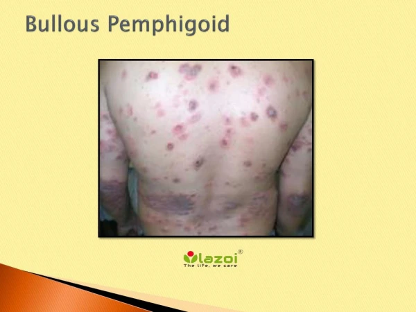
Bullous Pemphigoid • Elderly M+F • Subepidermal bullae often predated by itchy urticated areas on limb girdles • Tense blisters last several days •pruritus • Mucosal lesions unusual, patients well • good prognosis.
B.P. -Skin biopsy shows a deeper blister(than in pemphigus) owing to a subepidermal split through the BM -On direct IF, perilesional skin shows linear band of IgG and C3 along BMZ -Indirect IF shows IgG antibodies that reacts with the BMZ in most patients
B.P. treatment SteroidTopical Systemic Steroid Antibiotics : Tetracycline, Minocin Dapsone Immuno suppressive.