Download

1 / 40
480 likes | 988 Views
Evaluation of Mitral Stenosis. Echo Conference April 6, 2011 Frances Canet, MD. Outline. Causes and Anatomy Assessment of Mitral Stenosis How to Grade Mitral Stenosis Cases and Application. Causes and Anatomy. Rheumatic MS Commissural fusion Degenerative MS Annular calcification

E N D
Evaluation of Mitral Stenosis Echo Conference April 6, 2011 Frances Canet, MD
Outline • Causes and Anatomy • Assessment of Mitral Stenosis • How to Grade Mitral Stenosis • Cases and Application
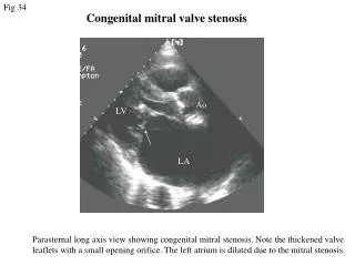
Causes and Anatomy Rheumatic MS Commissural fusion Degenerative MS Annular calcification Associated with elderly, hypertension, atherosclerosis and aortic stenosis Congenital MS Abnormalities of subvalvular apparatus Other: Systemic lupus, infiltrative disease, carcinoid heart disease, drug-induced valve disease
How to Assess Mitral Stenosis Level 1 Recommendations: Pressure gradient MVA Planimetry Pressure half-time Level 2 Recommendations: Continuity equation Proximal isovelocity surface area method (PISA) Stress echocardiography
Pressure Gradient Continuous wave doppler is preferred Gradient is measured in the apical window Color doppler is used to identify eccentric diastolic mitral jets Doppler beam is guided by the highest flow velocity zone identified by color doppler Mean gradient is the relevant hemodynamic finding Measure heart rate at which gradients are obtained If patient is in atrial fibrillation, the mean gradient should be an average of five cycles with the least variation of R-R intervals
Mitral Valve Area Planimetry • Direct tracing of the mitral orifice including opened commissures in the parasternal short-axis view at mid-diastole • Advantages: • Direct measure of MVA • Does not involve hypothesis regarding flow conditions, cardiac chamber compliance or associated valvular lesions • Best correlation with anatomic valve area of explanted valves
Mitral Valve Area Planimetry • Obtaining and measuring the image: • Scan apex to the base of the LV to ensure the cross-sectional area is measured at the leaflet tips. • Plane should be perpendicular to the mitral orifice, elliptical shape. • Gain, sufficient to see contour of the mitral orifice. • If too excessive, may cause under estimation of the valve area. • Perform several measurements if the patient has atrial fibrillation or incomplete commissural fusion
Pressure half-time T1/2 = time interval in milliseconds between the maximum mitral gradient in early diastole and the time point where the gradient is half the maximum initial value MVA = 220/T1/2
Measuring T1/2 with a bimodal, non-linear decreasing slope of the E-wave
Continuity equation – Level 2 Based on assumption that the filling volume of diastolic mitral flow is equal to aortic SV. MVA = pi (D2/4) (VTIAortic/ VTIMitral) D is the diameter of the LVOT in cm VTI is in cm. Accuracy and reproducibility is hampered by the number of measurements increasing the impact of errors of measurements. Cannot be used in atrial fibrillation or associated significant MR or AR
Proximal isovelocity surface area method – Level 2 MVA = pi (r2) (Valiasing) / Peak Vmitral x alpha/1800 R is the radius of the convergence hemisphere in cm Valiasing is the aliasing velocity in cm/s Peak Vmitralis the peak CWD velocity of mitral inflow in cm/s alpha is the opening angle of mitral leaflets relative to flow direction
Valve Anatomy Parasternal short-axis view valve thickness (maximum and heterogeneity) commissural fusion extension and location of localized bright zones (fibrous nodules or calcification) Parasternal long-axis view valve thickness extension of calcification valve pliability subvalvular apparatus (chordal thickening, fusion, or shortening) Apical two-chamber view subvalvular apparatus (chordal thickening, fusion, or shortening) Detail each component and summarize in a score
Stress Echocardiography – Level 2 Enables measurement of mean mitral gradient and systolic pulmonary artery pressure during effort. Semi-supine exercise echocardiography allows monitoring of gradient. Useful in patients with equivocal or discordant with the severity of MS.
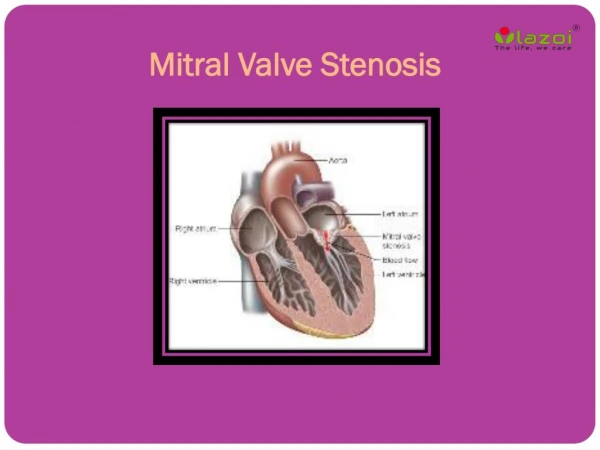
How to Grade Mitral Stenosis Normal MVA is 4.0-5.0 cm2 MVA >1.5 cm2 does not produce symptoms As severity increases, cardiac output decreases and fails to increase during exercise.
Wilkins (Valvotomy )Score • Grades morphological changes in the MV during echo: • Leaflet mobility • Leaflet thickening • Valve calcification • Involvement of the subvalvular apparatus • Each characteristic is graded from 0-4, with a total of 16 points total. • A score >8 is predictive of low success post percutaneous mitral valvuloplasty.
Case 1 • 72-year-old man with known moderate aortic stenosis, mitral regurgitation, hypertension, diabetes, COPD, TIA and severe pulmonary hypertension based on cardiac catheterization results is referred for echocardiogram to assess severity of mitral valve regurgitation. • How severe is his mitral regurgitation? Does he have mitral stenosis? What are his options for repair – calculate valvotomy score?
Continuity equation • LVOT Diameter is 2.1 • VTI aortic is 87 • VTI mitral is 87.2 • MVA = pi (D2/4) (VTIAortic / VTIMitral) • MVA = 3.89 cm2 (Not accurate compared to MVA of 1.15 cm2 calculated from pressure gradient. Remember, it is not accurate in patient with severe mitral regurgitation or atrial fibrillation.) • Less accurate calculation of MVA as it relies on several other measurements to be accurate.
Valvotomy Score = 12 Mobility – valve moves forward in diastole, moves mainly from base 3 points Subvalvular Thickening – thickening of chordal structures extending into 1/3rd of the chordal length 3 points Thickening – extends through the entire leaflet 3 points Calcification – Brightness extending into the mid-portion of the leaflets 3 points Total score = 12
Case 2 • 56-year-old woman with a history of rheumatic mitral valve stenosis, respiratory failure, heart failure, atrial fibrillation, recent stroke, COPD, sarcoidosis, schizophrenia was transferred from an outside hospital for a second opinion on mitral valve replacement. She has poor functional and neurologic status at present. • Evaluate the grade of her mitral stenosis and calculate her valvotomy score.
Planimetry Still This is not acutally the area of the MV orfiice. Look at the small sliver of black area just below the tracing.
Grade of mitral stenosis: Severe Resting mean pressure gradient: 16mmHg (severe is >10mmHg) Mitral valve area using half time: 0.77cm2 (severe is <1.0 cm2) PHT: 285 ms (severe is greater than 220ms)
Valvotomy score: 14 out of 16 Mobility: 4 – No or minimal forward movement of the leaflets. Subvalvular Thickening: 2-3- Thickening of chordal structures up to one-third of the chordal length possibly to distal third of the chords. Thickening: 4 – Considerable thickening of all leaflet tissue (>8-10mm). Calcification: 4 – Extensive brightness throughout much of the leaflet tissue.