Download
1 / 28
280 likes | 292 Views
The Special Senses Vision, Taste, Smell. 15. Eye and Associated Structures. 70% of all sensory receptors are in the eye Most of the eye is protected by a cushion of fat and the bony orbit

E N D
The Special Senses Vision, Taste, Smell 15
Eye and Associated Structures • 70% of all sensory receptors are in the eye • Most of the eye is protected by a cushion of fat and the bony orbit • Accessory structures include eyebrows, eyelids, conjunctiva, lacrimal apparatus, and extrinsic eye muscles
Eyebrows Eyelids • Coarse hairs that overlie the supraorbital margins • Functions include: • Shading the eye • Preventing perspiration from reaching the eye Eyelids Protect the eye anteriorly • Palpebral fissure – separates eyelids • Canthi – medial and lateral angles (commissures)
Palpebrae (Eyelids) • Eyelashes • Project from the free margin of each eyelid • Initiate reflex blinking • Lubricating glands associated with the eyelids • Meibomian glands and sebaceous glands • Ciliary glands lie between the hair follicles
Lacrimal Apparatus (tear ducts) Lacrimal gland and associated ducts • Lacrimal glands secrete tears • Tears • Contain mucus, antibodies, and lysozyme • Drain into the nasolacrimal duct
Extrinsic Eye Muscles • Six straplike extrinsic eye muscles • Enable the eye to follow moving objects • Maintain the shape of the eyeball
Structure of the Eyeball • A slightly irregular hollow sphere with anterior and posterior poles • The wall is composed of three tunics – fibrous, vascular, and sensory • The internal cavity is filled with fluids called humors • The lens separates the internal cavity into anterior and posterior segments
Structure of the Eyeball Figure 15.8a
Vascular Tunic: Iris • The colored part of the eye • Pupil – central opening of the iris • Regulates the amount of light entering the eye during: • Close vision and bright light – pupils constrict • Distant vision and dim light – pupils dilate • Changes in emotional state – pupils dilate when the subject matter is appealing or requires problem-solving skills
Pupil Dilation and Constriction Figure 15.9
Sensory Tunic: Retina • Two-layered membrane • Pigmented layer – the outer layer that absorbs light and prevents its scattering • Neural layer, which contains: • Photoreceptors • Bipolar cells and ganglion cells • Amacrine and horizontal cells
The Retina: Ganglion Cells and the Optic Disc • Ganglion cell axons: • Run along the inner surface of the retina • Leave the eye as the optic nerve • The optic disc: • Is the site where the optic nerve leaves the eye • Lacks photoreceptors (the blind spot)
The Retina: Photoreceptors • Rods: • Respond to dim light • Are used for peripheral vision • Cones: • Respond to bright light • Have high-acuity color vision • Are found in the macula lutea • Are concentrated in the fovea centralis
Channels open in the DARK- continuous release of neurotransmitter from photoreceptor- release of neurotransmitter from photoreceptor hyperpolarizes bipolar calcium channels to close- no neurotransmitter release Light- closes channels in photoreceptor/opens calcium channels in bipolar cell- neurotransmitter released- action potential in optic nerve
Chemical Senses • Chemical senses – gustation (taste) and olfaction (smell) • Their chemoreceptors respond to chemicals in aqueous solution • Taste – to substances dissolved in saliva • Smell – to substances dissolved in fluids of the nasal membranes
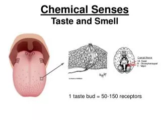
Taste Buds • There are five (maybe 6) basic taste sensations • Sweet – sugars, saccharin, alcohol, and some amino acids • Salt – metal ions • Sour – hydrogen ions • Bitter – alkaloids such as quinine and nicotine • Umami – elicited by the amino acid glutamate • Lipids? ? Most of the 10,000 or so taste buds are found on the tongue
Physiology of Taste • In order to be tasted, a chemical: • Must be dissolved in saliva • Must contact gustatory hairs • Binding of the food chemical: • Depolarizes the taste cell membrane, releasing neurotransmitter • Initiates a generator potential that elicits an action potential
Taste Transduction • The stimulus energy of taste is converted into a nerve impulse (action potential) by: • Na+ influx in salty tastes • H+ in sour tastes (by directly entering the cell, by opening cation channels, or by blockade of K+ channels) • Gustducin in sweet and bitter tastes
Gustatory Pathway Figure 15.2
Influence of Other Sensations on Taste • Taste is 80% smell • Thermoreceptors, mechanoreceptors, nociceptors also influence tastes • Temperature and texture enhance or detract from taste
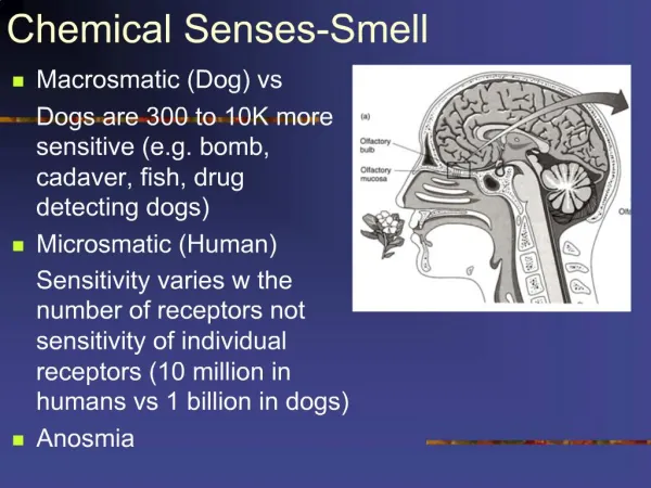
Sense of Smell • The organ of smell is the olfactory epithelium, which covers the superior nasal concha • Olfactory receptor cells are bipolar neurons with radiating olfactory cilia • Olfactory receptors are surrounded and cushioned by supporting cells • Basal cells lie at the base of the epithelium
Sense of Smell Figure 15.3
Physiology of Smell • Olfactory receptors respond to several different odor-causing chemicals • When bound to ligand these proteins initiate a G protein mechanism, which uses cAMP as a second messenger • cAMP opens Na+ and Ca2+ channels, causing depolarization of the receptor membrane that then triggers an action potential
Olfactory Pathway • Impulse sent to: • The olfactory cortex (what is that smell) • The hypothalamus, amygdala, and limbic system (emotional component of smell
Olfactory Transduction Process Na+ Odorant binding protein Odorant chemical Active Inactive Na+ influx causes depolarization ATP Adenylate cyclase cAMP Depolarization of olfactory receptor cell membrane triggers action potentials in axon of receptor Cytoplasm Figure 15.4