Download
1 / 20
200 likes | 276 Views
SPECIAL SENSES: VISION. MARTINI, FUNDAMENTALS OF ANATOMY & PHYSIOLOGY, 8 TH EDITION, CHAPTER # 17 Exercise # 21. NOTE:. THIS IS A STUDY GUIDE , NOT AN ALL INCLUSIVE REVIEW. THERE MIGHT BE THINGS NOT COVERED BY THIS STUDY GUIDE THAT MIGHT BE ASKED IN YOUR PRACTICUMS /

E N D
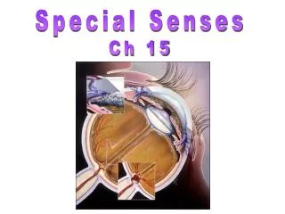
SPECIAL SENSES: VISION MARTINI, FUNDAMENTALS OF ANATOMY & PHYSIOLOGY, 8TH EDITION, CHAPTER # 17 Exercise # 21
NOTE: • THIS IS A STUDY GUIDE, NOT AN ALL INCLUSIVE REVIEW. • THERE MIGHT BE THINGS NOT COVERED BY THIS STUDY GUIDE THAT MIGHT BEASKED IN YOUR PRACTICUMS / QUIZZES. • STUDENTS ARE RESPONSIBLE FOR READING THEIR TEXBOOK (S) AND FOR ALL THE MATERIAL COVERED DURING THE LABORATORY PERIOD, AS PER THE COURSE SYLLABUS
OBJECTIVES • Identifying the structures of the eye and describe their functions.
VISION Accessory structures of the eye Eyeball Aqueous humor Scleral venous sinus Vitreous humor or body
ACCESSORY STRUCTURES: GLANDS • Lacrimal gland- tear production • Lacrimal sac- collect tears & send them to the nasolacrimal duct • Nasolacrimal duct- to drain excess of tears to the nasal cavity
EYEBALL: COATS • Cornea (clear outer part)- it allows the light to come in • Sclera(white part)- it protects & gives the shape to the eyes • Choroids - to nourish &to absorb excess of light • Ciliary body- to contain the ciliary muscle and ciliary process • Suspensory ligament- it holds the lens in place • Iris (part with color)- to control the size of pupil • Pupil (hole)- it lets the light to go inside
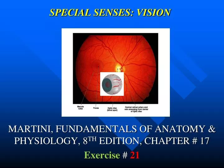
Retina- it contains photoreceptors • Ganglion cells (their axons form the optic • nerve)-they carry visual info • Bipolar cells-to connect the ganglion cells • to the photoreceptors • Photoreceptors- to capture the light • Rods- night, white & black vision • Cones- bright & color vision • Optic disc(blind spot)- area without • photoreceptors where the optic nerve goes out • Macula lutea- it contains high • concentration of cones (yellow spot) • Fovea centralis(inside the macula lutea)- • To give sharpest vision
Lens- to focus the light • Cavities • Anterior cavity- it contains the aqueous humor • Anterior chamber (space between the cornea • & Iris)- it contains aqueous humor • Posterior chamber (between iris & lens)- it also contains • aqueous humor • Posterior cavity(area behind the lens)- • it contains vitreous humor
AQUEOUS HUMOR, SCLERAL VENOUS SINUS & VITREOUS HUMOR • Aqueous humor- to nourish the lens & the cornea • Because they do not have blood vessels • Sclera venous sinus(canal of schelmm)- to drain • the aqueous humor. • Important- if obstruction = glaucoma = blindness • Vitreous humor- it maintains the shape of the • lens & prevents collapse
REMEMBER!GO TO THE TUTORING ROMMAND PRACTICE WITH MODELS.ROMM 3326.