Download
1 / 47
470 likes | 479 Views
Chapter 11: Cell Communication “ No man ’ s an island and neither is a cell. ”. Evolution of Cell Signaling. A signal-transduction pathway is a series of steps by which a signal on a cell ’ s surface is converted into a specific cellular response

E N D
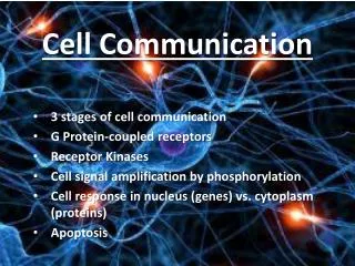
Chapter 11: Cell Communication “No man’s an island and neither is a cell.”
Evolution of Cell Signaling • A signal-transduction pathway is a series of steps by which a signal on a cell’s surface is converted into a specific cellular response • Signal transduction pathways convert signals on a cell’s surface into cellular responses • Pathway similarities suggest that ancestral signaling molecules evolved in prokaryotes and have since been adopted by eukaryotes
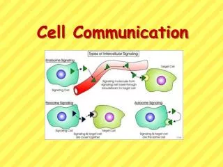
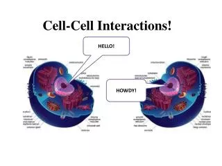
Local and Long-Distance Signaling • Cells in a multicellular organisms communicate by chemical messengers • Animal and plant cells have cell junctions that directly connect the cytoplasm of adjacent cells • In local signaling, animal cells may communicate by direct contact
LE11-3 Plasma membranes Plasmodesmata between plant cells Gap junctions between animal cells Cell junctions Cell-cell recognition
In many other cases, animal cells communicate using local regulators, messenger molecules that travel only short distances • In long-distance signaling, plants and animals use chemicals called hormones
LE 11-4 Local signaling Long-distance signaling Target cell Endocrine cell Blood vessel Electrical signal along nerve cell triggers release of neurotransmitter Neurotransmitter diffuses across synapse Secreting cell Secretory vesicle Hormone travels in bloodstream to target cells Local regulator diffuses through extracellular fluid Target cell Target cell is stimulated Paracrine signaling Synaptic signaling Hormonal signaling
The Three Stages of Cell Signaling: A Preview • Earl W. Sutherland discovered how the hormone epinephrine acts on cells • Sutherland suggested that cells receiving signals went through three processes: • Reception • Transduction • Response Animation: Overview of Cell Signaling
LE 11-5_1 EXTRACELLULAR FLUID CYTOPLASM Plasma membrane Reception Transduction Receptor Signal molecule
LE 11-5_2 EXTRACELLULAR FLUID CYTOPLASM Plasma membrane Reception Transduction Receptor Relay molecules in a signal transduction pathway Signal molecule
LE 11-5_3 EXTRACELLULAR FLUID CYTOPLASM Plasma membrane Reception Transduction Response Receptor Activation of cellular response Relay molecules in a signal transduction pathway Signal molecule
Concept 11.2: Reception: A signal molecule binds to a receptor protein, causing it to change shape • The binding between a signal molecule (ligand) and receptor is highly specific • A conformational change in a receptor is often the initial transduction of the signal • Most signal receptors are plasma membrane proteins
Intracellular Receptors • Some receptor proteins are intracellular, found in the cytosol or nucleus of target cells • Small or hydrophobic chemical messengers can readily cross the membrane and activate receptors • Examples of hydrophobic messengers are the steroid and thyroid hormones of animals • An activated hormone-receptor complex can act as a transcription factor, turning on specific genes
LE 11-6 Hormone (testosterone) EXTRACELLULAR FLUID The steroid hormone testosterone passes through the plasma membrane. Plasma membrane Testosterone binds to a receptor protein in the cytoplasm, activating it. Receptor protein Hormone- receptor complex The hormone- receptor complex enters the nucleus and binds to specific genes. DNA The bound protein stimulates the transcription of the gene into mRNA. mRNA NUCLEUS New protein The mRNA is translated into a specific protein. CYTOPLASM
Receptors in the Plasma Membrane • Most water-soluble signal molecules bind to specific sites on receptor proteins in the plasma membrane • There are three main types of membrane receptors: • G-protein-linked receptors • Receptor tyrosine kinases • Ion channel receptors
Transmembrane Receptor Structure • All three transmembrane receptor types have the same basic structural features: 1) an extracellular domain (ECD), 2) a transmembrane domain (TMD), and 3) an intracellular domain (ICD) ECD binds ligand (Extracellular Space) TMD anchors protein in the membrane (Cell Membrane) (Cytoplasm) ICD shape change occurs to relay internalization of signal
A G-protein-linked receptor is a plasma membrane receptor that works with the help of a G protein • The G-protein acts as an on/off switch: If GDP is bound to the G protein, the G protein is inactive
Receptor tyrosine kinases are membrane receptors that attach phosphates to tyrosines • A receptor tyrosine kinase can trigger multiple signal transduction pathways at once • Reminder: A kinase is a class of enzyme that phosphorylates its substrate
LE 11-7b Signal molecule Signal-binding site a Helix in the membrane Signal molecule Tyr Tyr Tyrosines Tyr Tyr Tyr Tyr Tyr Tyr Tyr Tyr Tyr Tyr Tyr Tyr Tyr Tyr Tyr Tyr Receptor tyrosine kinase proteins (inactive monomers) Dimer CYTOPLASM Activated relay proteins Cellular response 1 P P Tyr Tyr P Tyr Tyr Tyr Tyr P P P Tyr Tyr P Tyr Tyr Tyr Tyr P P P Cellular response 2 Tyr Tyr P Tyr Tyr Tyr P Tyr 6 ATP 6 ADP Activated tyrosine- kinase regions (unphosphorylated dimer) Fully activated receptor tyrosine-kinase (phosphorylated dimer) Inactive relay proteins
An ion channel receptor acts as a gate when the receptor changes shape • When a signal molecule binds as a ligand to the receptor, the gate allows specific ions, such as Na+ or Ca2+, through a channel in the receptor
LE 11-7c Gate closed Signal molecule (ligand) Ions Plasma membrane Ligand-gated ion channel receptor Gate open Cellular response Gate closed
Concept 11.3: Transduction: Cascades of molecular interactions relay signals from receptors to target molecules in the cell • Transduction usually involves multiple steps • Multistep pathways can amplify a signal: A few molecules can produce a large cellular response • Multistep pathways provide more opportunities for coordination and regulation
Signal Transduction Pathways • The molecules that relay a signal from receptor to response are mostly proteins • Like falling dominoes, the receptor activates another protein, which activates another, and so on, until the protein producing the response is activated https://www.youtube.com/watch?v=WRv9b7S6Hm8 • At each step, the signal is transduced into a different form, usually a conformational change
Protein Phosphorylation and Dephosphorylation • In many pathways, the signal is transmitted by a cascade of protein phosphorylations • Phosphatase enzymes remove the phosphates; these are the counterparts to kinases • This phosphorylation and dephosphorylation system acts as a molecular switch, turning activities on and off
LE 11-8 Signal molecule Receptor Activated relay molecule Inactive protein kinase 1 Active protein kinase 1 Inactive protein kinase 2 ATP ADP P Active protein kinase 2 Phosphorylation cascade PP P i Inactive protein kinase 3 ATP ADP P Active protein kinase 3 PP P i Inactive protein ATP P ADP Cellular response Active protein PP P i
Small Molecules and Ions as Second Messengers • Second messengers are small, nonprotein, water-soluble molecules or ions • The extracellular signal molecule that binds to the membrane is a pathway’s “first messenger” • Second messengers can readily spread throughout cells by diffusion • Second messengers participate in pathways initiated by G-protein-linked receptors and receptor tyrosine kinases
Cyclic AMP • Cyclic AMP (cAMP) is one of the most widely used second messengers • Adenylyl cyclase, an enzyme in the plasma membrane, converts ATP to cAMP in response to an extracellular signal
LE 11-9 Phosphodiesterase Adenylyl cyclase Pyrophosphate H2O P P i ATP Cyclic AMP AMP
Many signal molecules trigger formation of cAMP • Other components of cAMP pathways are G proteins, G-protein-linked receptors, and protein kinases • cAMP usually activates protein kinase A, which phosphorylates various other proteins • Further regulation of cell metabolism is provided by G-protein systems that inhibit adenylyl cyclase
LE 11-10 First messenger (signal molecule such as epinephrine) Adenylyl cyclase G protein G-protein-linked receptor GTP ATP Second messenger cAMP Protein kinase A Cellular responses
Calcium ions and Inositol Triphosphate (IP3) • Calcium ions (Ca2+) act as a second messenger in many pathways • Calcium is an important second messenger because cells can regulate its concentration
A signal relayed by a signal transduction pathway may trigger an increase in calcium in the cytosol • Pathways leading to the release of calcium involve inositol triphosphate (IP3) and diacylglycerol (DAG) as second messengers Animation: Signal Transduction Pathways
LE 11-12_1 EXTRACELLULAR FLUID Signal molecule (first messenger) G protein DAG GTP G-protein-linked receptor PIP2 Phospholipase C IP3 (second messenger) IP3-gated calcium channel Endoplasmic reticulum (ER) Ca2+ CYTOSOL
LE 11-12_2 EXTRACELLULAR FLUID Signal molecule (first messenger) G protein DAG GTP G-protein-linked receptor PIP2 Phospholipase C IP3 (second messenger) IP3-gated calcium channel Endoplasmic reticulum (ER) Ca2+ Ca2+ (second messenger) CYTOSOL
LE 11-12_3 EXTRACELLULAR FLUID Signal molecule (first messenger) G protein DAG GTP G-protein-linked receptor PIP2 Phospholipase C IP3 (second messenger) IP3-gated calcium channel Cellular re- sponses Various proteins activated Endoplasmic reticulum (ER) Ca2+ Ca2+ (second messenger) CYTOSOL
Concept 11.4: Response: Cell signaling leads to regulation of cytoplasmic activities or transcription • Ultimately, a signal transduction pathway leads to regulation of one or more cellular activities • The response may occur in the cytoplasm or may involve action in the nucleus • Many pathways regulate the activity of enzymes
Many other signaling pathways regulate the synthesis of enzymes or other proteins, usually by turning genes on or off in the nucleus • The final activated molecule may function as a transcription factor
LE 11-14 Growth factor Reception Receptor Phosphorylation cascade Transduction CYTOPLASM Inactive transcription factor Active transcription factor Response P DNA Gene NUCLEUS mRNA
Control of Cell Response: Fine-Tuning of the Response – Amplification & Specificity • Multistep pathways have two important benefits: 1. Signal Amplification: - Enzyme cascades amplify the cell’s response - At each step, the number of activated products is much greater than in the preceding step http://highered.mcgraw-hill.com/sites/0072437316/student_view0/chapter7/animations.html# 2. Contributing to the specificity of the response:
LE 11-13 Reception Binding of epinephrine to G-protein-linked receptor (1 molecule) Transduction Inactive G protein Active G protein (102 molecules) Inactive adenylyl cyclase Active adenylyl cyclase (102) ATP Cyclic AMP (104) Inactive protein kinase A Active protein kinase A (104) Inactive phosphorylase kinase Active phosphorylase kinase (105) Inactive glycogen phosphorylase Active glycogen phosphorylase (106) Response Glycogen Glucose-1-phosphate (108 molecules)
Different kinds of cells have different collections of proteins • These differences in proteins give each kind of cell specificity in detecting and responding to signals • The response of a cell to a signal depends on the cell’s particular collection of proteins • Pathway branching and “cross-talk” further help the cell coordinate incoming signals
LE 11-15 Signal molecule Receptor Relay molecules Response 2 Response 3 Response 1 Cell A. Pathway leads to a single response Cell B. Pathway branches, leading to two responses Same ligand may bind more than one type of receptor Activation or inhibition Response 5 Response 4 Cell D. Different receptor leads to a different response Cell C. Cross-talk occurs between two pathways
Control of the Cell Response: Signaling Efficiency – Scaffolding Proteins and Signaling Complexes • Scaffolding proteins are large relay proteins to which other relay proteins are attached • Scaffolding proteins can increase the signal transduction efficiency
LE 11-16 Signal molecule Plasma membrane Receptor Three different protein kinases Scaffolding protein
Control of Cell Response: Termination of the Signal • Inactivation mechanisms are an essential aspect of cell signaling • When signal molecules leave the receptor, the receptor reverts to its inactive state • Reception of another signal that inactivates the first pathway (phosphorylation/dephosphorylation) • Additionally, when certain receptors become modified, they can become internalized and ubiquitinated (tagged for degradation) - effectively terminating the cascade pathway
Concept 11.5: Apoptosis • Also known as programmed cell death (PCD) • Different from necrosis – cells don’t rupture (lyse), thus, cellular contents are not released to the surrounding environment
Caspases – the main proteases (enzymes that degrade proteins) • of apoptosis