Download
1 / 27
280 likes | 307 Views
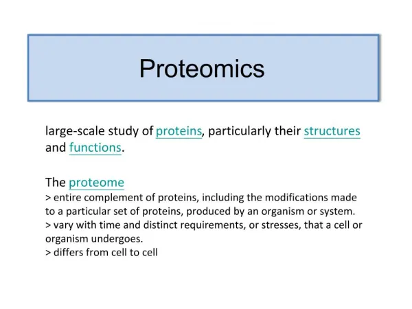
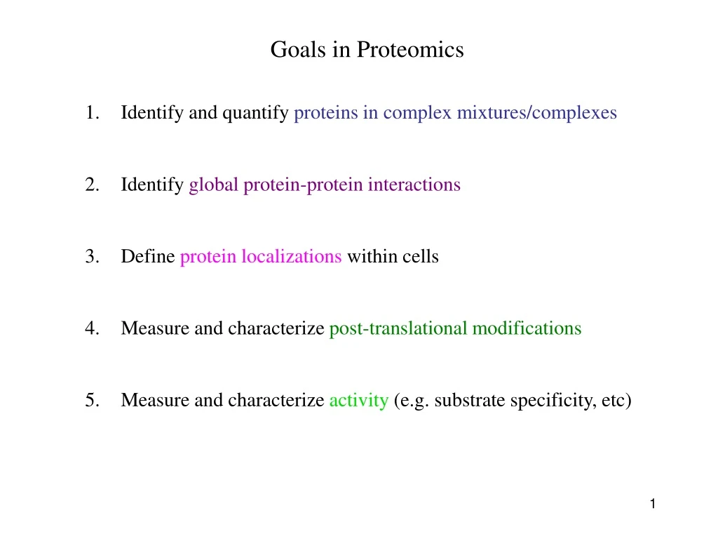
Goals in Proteomics. Identify and quantify proteins in complex mixtures/complexes Identify global protein-protein interactions Define protein localizations within cells 4. Measure and characterize post-translational modifications

E N D
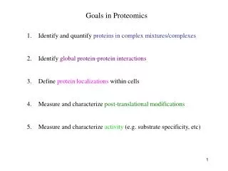
Goals in Proteomics • Identify and quantify proteins in complex mixtures/complexes • Identify global protein-protein interactions • Define protein localizations within cells 4. Measure and characterize post-translational modifications • Measure and characterize activity (e.g. substrate specificity, etc)
Goals in Proteomics • Identify and quantify proteins in complex mixtures/complexes MS and MS/MS • Identify global protein-protein interactions MS and MS/MS, Y2H • Define protein localizations within cells High-throughput microscopy, organelle pull down 4. Measure and characterize post-translational modifications MS techniques • Measure and characterize activity (e.g. substrate specificity, etc) Protein arrays
Basic overview of Tandem mass-spectrometry (MS/MS) Coon et al. 2005
Intro to Mass Spec (MS) Separate and identify peptide fragments by their Mass and Charge (m/z ratio) Mass Spec MS Spectrum Ion source Mass analyzer Detector Basic principles: 1. Ionize (i.e. charge) peptide fragments 2. Separate ions by mass/charge (m/z) ratio 3. Detect ions of different m/z ratio 4. Compare to database of predicted m/z fragments for each genome
Intro to Mass Spec (MS) Separate and identify peptide fragments by their Mass and Charge (m/z ratio) Ionization Goal: ionize (i.e. charge) peptide fragments without destroying molecule Positive ionization (protonate amine groups) especially useful for trypsinized proteins (cleaved after R and K) vs. Negative ionization (deprotonate carboxylics and alcohols) http://www.colorado.edu/chemistry/chem5181/MS_ESI_Gilman_Mashburn.pdf
Liquid chromatography + Electrospray ionization electric field * Commonly used with liquid solutions, more sensitive to contaminants, used for complex mixtures
Liquid chromatography + Electrospray ionization electric field * Commonly used with liquid solutions, more sensitive to contaminants, used for complex mixtures MALDI * Less sensitive to contaminants, more common for less complex mixtures http://www.astbury.leeds.ac.uk/facil/MStut/mstutorial.htm
Intro to Mass Spec (MS) Separate and identify peptide fragments by their Mass and Charge (m/z ratio) Ionization Goal: ionize (i.e. charge) peptide fragments without destroying molecule 2. Separation of ions based on m/z ratio (mass m versus charge c) Multiple flavors of mass analyzers use different technology. Different mass analyzers can be hooked together for unique properties.
Multiple flavors of mass analyzers Single MS (peptide fingerprinting): Identifies m/z of peptide only Peptide id’d by comparison to database, of predicted m/z of trypsinized proteins Tandem MS/MS (peptide sequencing): Pulls each peptide from the first MS Breaks up peptide bond Identifies each fragment based on m/z Collision cell
Multiple flavors of mass analyzers … can be hooked together in multiple configs. g. Orbitrap
Multiple flavors of mass analyzers Single MS (peptide fingerprinting): Identifies m/z of peptide only Peptide id’d by comparison to database, of predicted m/z of trypsinized proteins Tandem MS/MS (peptide sequencing): Pulls each peptide from the first MS Breaks up peptide bond Identifies each fragment based on m/z Collision cell Now multiple types of collision cells:CID: collision induced dissociation ETD: electron transfer dissociation HCD: high-energy collision dissociation
Fragmentation happens in fairly defined way along peptide backbone Peptide can fragment along 3 possible bonds … charge stays on either the Nt (a,b, or c) or Ct (x, y, or z) side of cleavagee With CID, cleavage along the CO-NH bond is most common, generating ‘b’ and ‘y’ ions * But phospho-modifications are labile via CID, and thus often missed
Electron-transfer dissociation (ETD) targets different bonds ETD cleavages favors ‘c’ and ‘z’ ions, is less sensitive to peptide sequence or length, and does not target phospho bonds
MS spectrum (i.e. peptide ions) Each peak often surrounded by smaller peaks of similar m/z Sensitivity of instrument determines resolution Each peak is a different peptide, separated based on m/z A single peptide is selected by the instrument for the second MS Mann Nat Reviews MBC. 5:699:711
Second MS identifies y (or b) ions to read out amino-acid sequence Mann Nat Reviews MBC. 5:699:711
Trypsin often used to digest proteins (cleaves after Arg and Lys) • Because of challenges distinguishing spectra, simplified mixtures • are typically injected into the MS: • either excised proteins • purified complexes • fractionated pools of complex mixtures
2 dimensional gel separation(largely outdated) The first dimension (separation by isoelectric focusing) - gel with an immobilised pH gradient - electric current causes charged proteins to move until it reaches the isoelectric point (pH gradient makes the net charge 0) The second dimension (separation by mass) -pH gel strip is loaded onto a SDS gel -SDS denatures the protein (to make movement solely dependent on mass, not shape) and eliminates charge. Ahna Skop
TAP-tag: Tandem Affinity Purification (for IP’ing individual proteins and proteins bound to them)
Ion exchange chromatography Anion exchange: Column is postively charged (can bind negatively charged proteins). Cation exchange: Column is negatively charged (can bind positively charged proteins). Exploit the isoelectric point of a protein to Separate it from other macromolecules. Ahna Skop
Affinity chromatography A ligand with high affinity to the protein is attached to a matrix. Protein of interest binds to ligand And is retained by resin. Everything else flows through. Can use excess of the soluble ligand to elute the protein. Ahna Skop
Phospho-proteomics and Post-translational modifications (PTMs) Phosphorylated (P’d) peptides are enriched, typically through chromatography - P’d peptides do not ionize as well as unP’d peptides - enrichment of P’d peptides ensures ionization and aids in mapping IMAC: immobilized metal ion affinity chromatography - phospho groups bind charged metals - contamination by negatively-charged peptides Titanium dioxide (TiO2) column: - binds phospho groups (mono-P’d better than multi-P’d) SIMAC: Sequential Elution from IMAC: - IMAC followed by TiO2 column Goal: identify which residues are phosphorylated (Ser, Thr, Tyr), mapped based on known m/z of phospho group
Quantitative proteomics Either absolute measurements or relatively comparisons • Spectral counting • Isotope labeling (SILAC) • Isobaric tagging (iTRAQ & TMT) • SRM
Spectral counting counting the number of peptides and counts for each protein Challenges: - different peptides are more (or less) likely to be assayed - analysis of complex mixtures often not saturating – may miss some peptides in some runs newer high-mass accuracy machines alleviate these challenges - quantitation comes in comparing separate mass-spec runs … therefore normalization is critical and can be confounded by error - requires careful statistics to account for differences in: quality of run, likelihood of observing each peptide, likelihood of observing each protein (eg. based on length, solubility, etc) Advantages / Challenges + label-free quantitation; cells can be grown in any medium - requires careful statistics to quantify - subject to run-to-run variation / error
SILAC (Stable Isotope Labeling with Amino acids in Cell culture) Cells are grown separately in heavy (13C) or light (12C) amino acids (often K or R), lysates are mixed, then analyzed in the same mass-spec run Mass shift of one neutron allows deconvolution, and quantification, of peaks in the same run. Advantages / Challenges: + not affected by run-to-run variation - need special media to incorporate heavy aa’s, - can only compare (and quantify) few samples directly - incomplete label incorporation can confound MS/MS identification
Isobaric Tagging iTRAQ or Tandem Mass Tags, TMTs Each peptide mix covalently tagged with one of 4, 6, or 8 chemical tags of identical mass Samples are then pooled and analyzed in the same MS run Collision before MS2 breaks tags – Tags can be distinguished in the small-mass range and quantified to give relative abundance across up to 8 samples. LTQ Velos Orbitrap Advantages / Challenges: + can analyze up to 8 samples, same run - still need to deal with normalization
Selective Reaction Monitoring (SRM) Targeted proteomics to quantify specific peptides with great accuracy • Specialized instrument capable of very sensitively measuring • the transition of precursor peptide and one peptide fragment • Typically dope in heavy-labeled synthetic peptides of precisely known • abundance to quantify Advantages: - best precision measurements Disadvantages: - need to identify ‘proteotypic’ peptides for doping controls - expensive to make many heavy peptides of precise abundance - limited number of proteins that can be analyzed
How does each spectrum translate to amino acid sequence? Mann Nat Reviews MBC. 5:699:711