Download
1 / 23
320 likes | 799 Views
Angiogenesis. Key features of angiogenesis. Tumour growth is angiogenesis-dependent Microvascular endothelial cells (ECs) are genetically-stable ECs release angiogenic factors that stimulate proliferation. Composition of nascent and mature blood vessel walls.

E N D
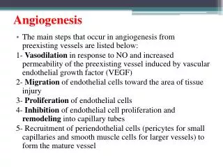
Key features of angiogenesis • Tumour growth is angiogenesis-dependent • Microvascular endothelial cells (ECs) are genetically-stable • ECs release angiogenic factors that stimulate proliferation
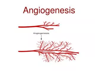
Composition of nascent and mature blood vessel walls • Nascent vessels consist of a tube of ECs, which mature into specialized capillaries, arteries and veins. • (b) Capillaries consist of ECs surrounded by basement membrane and a sparse layer of pericytes embedded within the EC basement membrane. Capillary endothelial layer can be continuous (muscle), fenestrated (kidney/ endocrine glands) or discontinuous (liver sinusoids). The endothelia of the blood-brain barrier or blood-retina barrier are further specialized to include tight junctions, and are thus impermeable to various molecules. • (c) Arterioles and venules have an increased coverage of mural cells compared with capillaries.
Steps in network formation and maturation during embryonic (physiological) angiogenesis
Key differences in tumour vasculature Different flow characteristics or blood volume Microvasculature permeability Increased fractional volume of extravascular, extracellular space
Steps in network formation and maturation during tumour angiogenesis
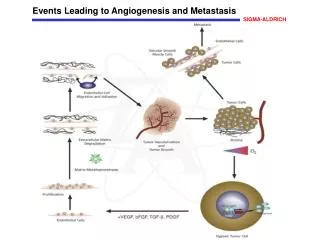
Cellular mechanisms of tumour angiogenesis 1 3 (1) host vascular network expands by budding of endothelial sprouts or formation of bridges (angiogenesis); (2) tumour vessels remodel and expand by the insertion of interstitial tissue columns into the lumen of pre-existing vessels (intussusception); and (3) endothelial cell precursors (angioblasts) home from the bone marrow or peripheral blood into tumours and contribute to the endothelial lining of tumour vessels (vasculogenesis) (4) Lymphatic vessels around tumours drain the interstitial fluid and provide a gateway for metastasizing tumour cells. 2 2 1 3 4 4
Cellular angiogenesis-overview Nature Reviews Drug Discovery1, 415-426 (2002)
Cellular angiogenesis-overview • Balance between inhibitory factors and angiogenic factors • Inhibitory – endostatin, angiostatin, thrombospondin • Angiogenic - VEGF, bFGF, PDGF • Tumour cells release pro-angiogenic factors which activate receptors (VEGFR) • also stimulates secretion and activation of MMPs which degrade the basement membrane • This allows activated endothelial cells (ECs) to migrate towards tumour, helped by integrins • ECs deposit a new basement membrane and secrete growth factors such as platelet-derived growth factor (PDGF), which attract supporting cells to stabilize the new vessel. VEGF – Vascular Endothelial Growth Factor bFGF - basic Fibroblast Growth Factor MMPs – Matrix MetalloProteinases PDGF – Platelet-Derived Growth Factor
4 2 1 3 5 Red: E-Cadherins Green: Integrins 6 Invasion & Metastasis
Integrins – the ‘velcro’ of the cell • The cell moves by "ruffling" it's membrane. This is done by a series of actin fibers, whose function is controlled by the integrins. These fibers cause the cell membrane to move in certain directions, and the integrins attach to the matrix as this happens, pulling the cell along a micrometer at a time
Red: E-Cadherins Green: Integrins Invasion & Metastasis
Epithelial-mesenchymal transition (EMT) necessary for invasiveness
Then why do secondary tumours histopathologically resemble primary tumours? EMT induced by stromal signals EMT may be reversible depending on the stromal signals e.g. TGF-b, TNF-a, EGF, HGF, IGF-1.
Cell invasiveness controlled by Matrix Metalloproteinases (MMPs) • MMPs secreted by stromal cells • Can be PM-bound or soluble enzymes • MMP activation can be indirect E.g. via urokinase plasminogen activator (uPA)
Cell motility regulated by RhoGTPases • Binary switches like Ras • 3 sub families; Rho, Rac and cdc42 Lysophosphatidic acid Overexpressing Rac Serum-starved GEF
Colonisation depends on a variety of factorsmetastatic tropisms (Paget’s ‘seed & soil’theory)
Colonisation depends on complex interactions between metastasising cells and their microenvironments E.g Osteolytic metastasis initiated by breast cancer
Bone growth versus loss Breast cancer initiated osteolytic metastasis
Reading Chapter 13 and 14 : Biology of Cancer by R Weinberg AND /OR Chapter 12: Cancer Biology by RJB King Angiogenesis in cancer and other diseases by P Carmeliet & RK. Jain Nature vol 407 14 september 2000 pp 249