Download
1 / 41
1.44k likes | 4.72k Views
Angiogenesis. CONTENTS. MECHANISM. PATHOLOGY EFFECT. TYPE. DEFINITION. DEFINITION. What is angiogenesis. Angiogenesis is the formation of new blood vessels from pre-existing vessels. Angiogenesis is a normal process in growth and development, as well as in wound healing.

E N D
CONTENTS MECHANISM PATHOLOGY EFFECT TYPE DEFINITION
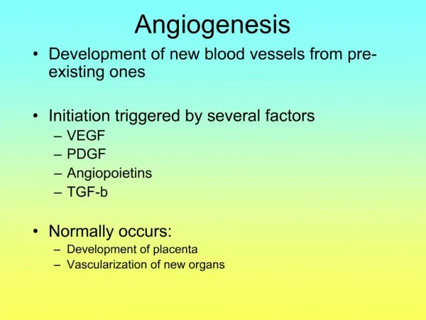
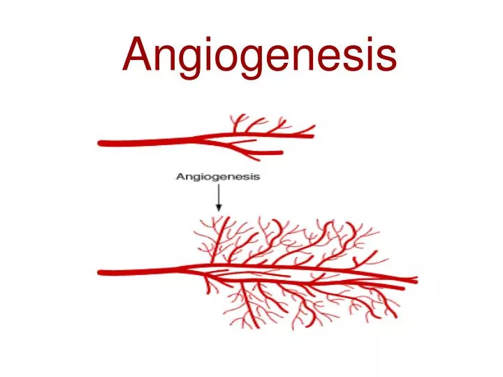
What is angiogenesis Angiogenesis is the formation of new blood vessels from pre-existing vessels. Angiogenesis is a normal process in growth and development, as well as in wound healing. However, this is also a fundamental step in the transition of tumors from a dormant state to a malignant state.
Angiogenesis is a process controlled by certain chemicals produced in the body. Some of these chemicals stimulate cells to repair damaged blood vessels or form new ones. Other chemicals, called angiogenesis inhibitors, signal the process to stop.
The balance hypothesis of the 'angiogenic switch'. Angiogenesis is tightly controlled by the balance of two sets of counteracting factors – angiogenic activators and inhibitors.
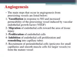
The angiogenic process The angiogenic process is a critical process for new cell and tissue growth。 The 4 major steps of endothelial cells in angiogenesis 1. Breaking through of the basal lamina that envelopes existing blood vessels 2. Migration toward a source signal 3. Proliferation 4. Formation of tubes Like most processes in homeostatic cellular systems, angiogenesis is a complex, highly regulated system. A large number of pro-angiogenic growth factors have been identified, many of which are capable of inducing all 4 of the above steps. One of the primary factors among these is a protein known as VEGF.
TYPE Sprouting angiogenesis Intussusceptive angiogenesis
Sprouting angiogenesis Sprouting angiogenesis was the first identified form of angiogenesis. It occurs in several well-characterized stages. First, biological signals known as angiogenic growth factors activate receptors present on endothelial cells present in pre-existing veins. Second, the activated endothelial cells begin to release enzymes called proteases that degrade the basement membrane in order to allow endothelial cells to escape from the original (parent) vessel walls.
The endothelial cells then proliferate into the surrounding matrix and form solid sprouts connecting neighboring vessels. As sprouts extend toward the source of the angiogenic stimulus, endothelial cells migrate in tandem, using adhesion molecules, the equivalent of cellular grappling hooks, called integrins. These sprouts then form loops to become a full-fledged vessel lumen as cells migrate to the site of angiogenesis. Sprouting occurs at a rate of several millimeters per day, and enables new vessels to grow across gaps in the vasculature. It is markedly different from splitting angiogenesis, however, because it forms entirely new vessels as opposed to splitting existing vessels.
Intussusceptive Angiogenesis Intussusception, also known as splitting angiogenesis, was first observed in neonatal rats. In this type of vessel formation, the capillary wall extends into the lumen to split a single vessel in two. There are four phases of intussusceptive angiogenesis. First, the two opposing capillary walls establish a zone of contact. Second, the endothelial cell junctions are reorganized and the vessel bilayer is perforated to allow growth factors and cells to penetrate into the lumen. Third, a core is formed between the two new vessels at the zone of contact that is filled with pericytes and myofibroblasts. These cells begin laying collagen fibers into the core to provide an extracellular matrix for growth of the vessel lumen. Finally, the core is fleshed out with no alterations to the basic structure.
Intussusceptive Angiogenesis Intussusception is important because it is a reorganization of existing cells. It allows a vast increase in the number of capillaries without a corresponding increase in the number of endothelial cells. This is especially important in embryonic development as there are not enough resources to create a rich microvasculature with new cells every time a new vessel develops.
The diagram of the difference between two kinds of angiogenesis
Different phases of embryonic vascular development. Intussusceptive angiogenesis Sprouting angiogenesis Endothelial precursors (angioblasts) differentiate to early endothelial cells (phase 1), which become assembled into a primitive capillary plexus (vasculogenesis) (phase 2). This emerging network expands via intussusceptive growth, intercalated growth and sprouting (angiogenesis) (phase 3), after which it becomes remodelled via pruning, fusion and regression of pre-existing vessels into a tree of arteries, capillaries and veins (phase 4).
Vasculogenesis Angiogenesis Arteriogenesis Modern terminology of angiogenesis Besides the differentiation between “Sprouting angiogenesis” and “Intussusceptive angiogenesis” today there exists the more common differentiation between the following types of angiogenesis .
Modern terminology of angiogenesis Vasculogenesis – Formation of vascular structures from circulating or tissue-resident endothelial stem cells(angioblasts), which proliferate into de novo endothelial cells. This form particularly relates to the embryonal development of the vascular system. Angiogenesis – Formation of thin-walled endothelium-lined structures with /without muscular smooth muscle wall and pericytes (fibrocytes). This form plays an important role during the adult life span, also as "repair mechanism" of damaged tissues. Arteriogenesis – Formation of medium-sized blood vessels possessing tunica media plus adventitia. Because it turned out that even this differentiation is not a sharp one, today quite often the term “Angiogenesis” is used summarizing all different types and modifications of arterial vessel growth.
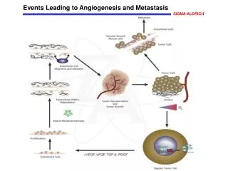
The relationship between tumor and angiogenesis While there are more than 100 distinct types of cancer (and considerable heterogeneity within each tumor type), there exists a remarkable similarity in the pathologic traits that collectively drive tumor growth. Across most—if not all—malignancies, sustained angiogenesis is considered to be one of these central hallmarks of cancer.
Angiogenesis is 1 of the 6 cellular transformations that lead to malignant growth The establishment of “sustained angiogenesis” as one of the fundamental “hallmarks of cancer” is based on more than a century of research.
Angiogenesis ? Tumor VEGF
Angiogenesis is a vital process in the progression of cancer from small, localized neoplasms to larger, growing, and potentially metastatic tumors. To grow beyond 1 to 2 mm in diameter, a tumor needs an independent blood supply, which is acquired by expressing growth factors that recruit new vasculature from existing blood vessels. This process continues even as the tumor matures. Thus, upregulation of angiogenesis is a key step in sustained tumor growth and may also be critical for tumor metastasis
Angiogenesis has been correlated with disease progression and/or poor prognosis in many tumor types—including lung, colon, breast, renal, and other cancers—and can be activated at different stages of tumor development, depending on the tumor type and microenvironmental conditions.
Angiogenesis is essential to tumor development http://images.google.co.kr/imgres?imgurl=http://www.researchvegf.com/researchvegf/images/Angiogenesis-tumor-growth.jpg&imgrefurl=http://www.researchvegf.com/researchvegf/ligand/hallmark/tumor-growth/index.m&usg=__Kin2MPD2XyIp91AtX3KVG-n4GRQ=&h=433&w=500&sz=66&hl=zh-CN&start=82&um=1&tbnid=KIaCa6YZDcAANM:&tbnh=113&tbnw=130&prev=/images%3Fq%3DAngiogenesis%26ndsp%3D21%26hl%3Dzh-CN%26lr%3D%26sa%3DN%26start%3D63%26um%3D1%26newwindow%3D1
VEGF: the predominant mediator of angiogenesis Among the many factors implicated in angiogenesis, VEGF has been identified as the most potent and predominant. The scope of scientific research involving VEGF continues to grow exponentially. From 1995 to 2005, the number of VEGF-related abstracts presented at the annual meeting of the American Society of Clinical Oncology (ASCO) increased 50-fold, highlighting the increased focus in research upon VEGF's role in oncology.
VEGF VEGF (also known as VEGF-A, but commonly referred to simply as VEGF) stands for “vascular endothelial growth factor.” This protein plays an important role in angiogenesis. As its name suggests, VEGF stimulates vascular endothelial cell growth, survival, and proliferation. As seen in preclinical models, VEGF has been shown to facilitate survival of existing vessels, contribute to vascular abnormalities (eg, tortuousness and hyperpermeability) that may impede effective delivery of antitumor compounds, and stimulate new vessel growth
The VEGF family of proteins VEGF is a member of a family of 6 structurally related proteins (see table below) that regulate the growth and differentiation of multiple components of the vascular system, especially blood and lymph vessels. The angiogenic effects of the VEGF family are thought to be primarily mediated through the interaction of VEGF with VEGFR-2. There are 4 major isoforms of VEGFA (VEGF), each coded for by a different portion of the VEGF gene. These isoforms are VEGF121, VEGF165, VEGF189, and VEGF206. Although these isoforms behave identically in solution, they differ in their ability to bind heparin and the extracellular matrix
RECEPTORS OF VEGF GROWTH FACTORS Three receptors have been identified that bind different VEGF growth factors: VEGFR1 (FLT1), VEGFR2 (Flk1/KDR), and VEGFR3 (FLT4) (initial receptor names are given in parentheses) These receptors belong to the superfamily of receptor tyrosine kinases (RTK) and, based on their structural peculiarities, they comprise a special class within it. Like all RTK, the VEGF receptors are transmembrane proteins with a single transmembrane domain . The extracellular region of VEGFR is formed by seven immunoglobulin-like domains (IG I-VII), whereas the intracellular part exhibits tyrosine kinase activity, and the tyrosine kinase domain in these receptors is separated to two fragments (TK-1 and TK-2) by an inter-kinase insert All VEGFR receptors are highly homologous .
Left: A tumor (tan) produces growth factors (yellow) that partner with receptors (orange) on blood vessels. This partnership stimulates the growth of new, though abnormal, blood vessels to nourish the tumor. Research suggests that little chemotherapy (green) can make its way through these defective blood vessels to poison the tumor. Right: The drug Avastin (blue) blocks growth factors from turning on their receptors. Research suggests that some of the faulty blood vessels begin to die off. Others form in a more orderly structure, which as a result improves both the delivery and the effectiveness of chemotherapy.