Download
1 / 1
10 likes | 100 Views

E N D
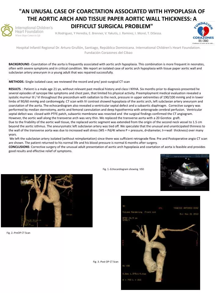
BACKGROUND.-Coarctationof the aorta is frequently associated with aortic arch hypoplasia. This combination is more frequent in neonates, often with severe symptoms and in critical condition. We report an isolated case of aortic arch hypoplasia with tissue paper aortic wall and subclavian artery aneurysm in a young adult that was repaired successfully. . METHODS.-Single isolated case; we reviewed the record and pre/ post-surgical CT scan RESULTS. : Patient is a male age 21 yo, without relevant past medical history and class I NYHA. Six months prior to diagnosis presented he several episodes of syncope like symptoms and chest pain, that limited his physical activity. Preemployment medical evaluation revealed a systolic murmur III / VI throughout the precordium with radiation to the neck, pressure in upper extremities of 190/100 mmHg and in lower limbs of 90/60 mmHg and cardiomegaly. CT scan with IV contrast showed hypoplasia of the aortic arch, left subclavian artery aneurysm and coarctationof the aorta. The echocardiogram also revealed a ventricular septal defect and a subaortic diaphragm. Corrective surgery was performed by median sternotomy, aortic and femoral cannulation and deep hypothermia with anterograde cerebral perfusion. Ventricular septal defect was closed with PTFE patch, subaortic membrane was resected and the surgical findings confirmed the CT angiogram. However, the aortic wall along the transverse arch was very thin. We replaced the transverse aorta with a 20 Goretex graft.Due to the friability of the aortic wall tissue, the replaced aortic segment was extended from the origin of the second neck vessel to 1.5 cm beyond the aortic isthmus. The aneurysmaticleft subclavian artery was tied off. We speculate that the unusual and unanticipated thinness to the wall of the transverse aorta was due to increased wall stress (WS = Pd/4t where P = pressure, d=diameter, t==wall thickness) over many years. We left the subclavian artery isolated (without reimplantation) since there was sufficient retrograde flow. Pre and Postoperative angio CT scan are shown. The patient returned to his normal life and his blood pressure is normal 6 months after surgery. CONCLUSIONS: Corrective surgery of the unusual adult presentation of aortic arch hypoplasia and coartationof aorta is feasible and provides good results and effective relief of symptoms. "An unusal case of coarctation associated with hypoplasia of the aortic arch and tissue paper aortic wall thickness: a difficult surgical problem" H.Rodriguez, Y Heredia, E. Brenner, V. Yakutis, J. Ramirez, I. Morel, T. DiSessa. Hospital Infantil Regional Dr. Arturo Grullón, Santiago, RepúblicaDominicana. International Children’s Heart Foundation. Fundación Corazones del Cibao Fig. 1.-Echocardiogram showing VSD. Fig. 2.-PreOP CT Scan Fig. 3.-Post OP CT Scan