Download
1 / 6
80 likes | 241 Views
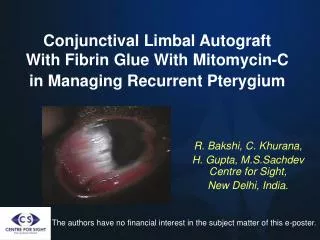
Amniotic Membrane as an Antifibrotic Agent in the Subconjunctival Space Surrounding a Conjunctival Autograft in Pterygium Surgery. John A. Hovanesian, M.D., Andrew Behesnilian Clinical Instructor, UCLA Jules Stein Eye Institute Harvard Eye Associates, Laguna Hills, California.

E N D
Amniotic Membrane as an Antifibrotic Agent in the Subconjunctival Space Surrounding a Conjunctival Autograft in Pterygium Surgery John A. Hovanesian, M.D., Andrew Behesnilian Clinical Instructor, UCLA Jules Stein Eye Institute Harvard Eye Associates, Laguna Hills, California Financial Interest Disclosure: Allergan, Abbott Medical Optics, Bausch & Lomb, Baxter BioScience, Diopter, Inc., Essex Woodlands Health Ventures, Inspire Pharmaceuticals, IOP, Inc, Ista Pharmaceuticals, Ocular Therapeutix, Revision optics, Sirion Therapeutics, Transcend Medical, True Vision Systems, Visiogen, Vista Research, Vistakon
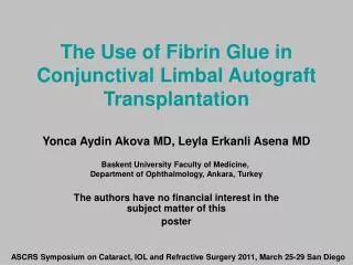
Pterygium surgery using a conjunctival autograft is associated with recurrence in 5% of cases. Recurrence arises from the conjunctival tissue surrounding the autograft. Amnionic membrane transplantation (AMT) has been used in place of the conjunctival autograft , but recurrence rates are similar to those with autografts. This is possibly because the amnion wears away from the ocular surface within 2-3 weeks of surgery. Introduction and Rationale amnion • We propose a method where AMT is used adjunctively as a biologic implant underneath the conjunctiva surrounding the autograft and not beneath the autograft. In this location protected from the ocular surface, the AMT may provide more prolonged anti-fibrotic effects.
We reviewed records of all patients who underwent this procedure in our office over a 19 month period Procedures were performed by a single surgeon (JAH). Postop Management Patch/shield overnight Drops Pred Forte QID Zymar QID First follow-up 1 week Methods
A conjunctival autograft is fashioned and left attached at the superior limbus while a graft of amniotic membrane is cut into a “c” shape and tucked into the subconjunctival space surrounding the site of pterygium excision. After this graft of amnion is fully placed in the subconjunctival space, the superior limbal graft is cut free and secured over the conjunctival defect with fibrin tissue adhesive.
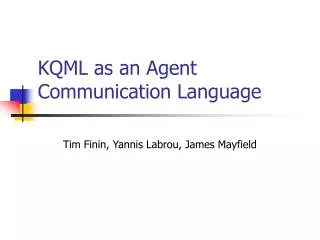
Results preop • 98 eyes of 94 patients • Mean follow-up9months (range 6-19) • All grafts remained viable and in place. • Postop appearance and inflammation was similar to standard conjunctival autograft. • Nasal conjunctival graft retraction occurred in 19 eyes but did not require re-intervention. • One pyogenicgranuloma occurred at the nasal edge of one graft. This resolved spontaneously. • No recurrences occurred in any eyes. two week postop
Conclusions Pterygium excision using combined conjunctival autograft and adjacent subconjunctival amnionic membrane transplantation is a viable technique for treatment of pterygium. Results compare favorably with established techniques. • John A. Hovanesian, M.D. • (949) 951-2020 • drhovanesian@harvardeye.com • More information on this technique available at www.bettereyesurgery.com