Download
1 / 11
180 likes | 299 Views
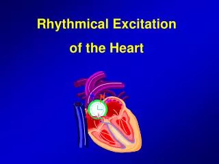
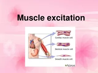
Rhythmical Excitation of the Heart. Arsalan Yousuf BS 4 th Semester. The rhythmical electrical impulses in a normal heart allows: The atria to contract about one sixth of a second ahead of ventricular contraction.

E N D
Rhythmical Excitation of the Heart Arsalan Yousuf BS 4th Semester
The rhythmical electrical impulses in a normal heart allows: • The atria to contract about one sixth of a second ahead of ventricular contraction. • Allows ventricular filling before they pump the blood through the lungs and peripheral circulation. • Allows all portions of the ventricles to contract almost simultaneously.
Sinus (Sinoatrial Node) Strip of specialized cardiac muscle Sinus nodal fibers connect directly with the atrial muscle fibers. Have the capability of self-excitation, a process that can cause automatic rhythmical discharge and contraction
Mechanism of Sinus Nodal Rhythmicity. The inherent leakiness of the sinus nodal fibers to sodium and calcium ions causes their self-excitation. • R.M.P of the sinus nodal fiber has a negativity of about -55 to -60 millivolts, in comparison with -85 to -90 millivolts for the ventricular muscle fiber. • Cell membranes of the sinus fibers are naturally leaky to sodium and calcium ions. • At -55 mV, the fast sodium channels already become “inactivated”. • The atrial nodal action potential is slower to develop than the action potential of the ventricular muscle.
Transmission of the Cardiac Impulse Through the Atria • The A-V node is located in the posterior wall of the right atrium immediately behind the tricuspid valve • Action potentials originating in the sinus node travel outward into the atrial muscle fibers and to the A-V node. • Impulse, after traveling through the internodal pathways, reaches the A-V node about 0.03 second after its origin in the sinus node. One-Way Conduction Through the A-V Bundle.
The impulse is delayed more than 0.1 second in the A-V nodal region before appearing in the ventricular septal A-V bundle. • Once it has entered this bundle, it spreads very rapidly through the Purkinje fibers to the entire endocardial surfaces of the ventricles. • Then the impulse once again spreads slightly less rapidly through the ventricular muscle to the epicardial surfaces.
Transmission in the Ventricular Purkinje System Special Purkinje fibers lead from the A-V node through the A-V bundle into the ventricles. Diminished numbers of gap junctions between successive cells in the conducting pathways allowing resistance to conduction of excitatory ions from one conducting fiber to the next. Distal portion of the A-V bundle passes downward in the ventricular septum for 5 to 15 millimeters toward the apex of the heart. Transmit action potentials at a velocity of 1.5 to 4.0 m/sec
Why Sinus node controls heart rhythymicity • The discharge rate of the sinus node is considerably faster than the natural self-excitatory discharge rate of either the A-V node or the Purkinje fibers. • Under abnormal conditions, few other parts of the heart can exhibit intrinsic rhythmical excitation in the same way like the sinus nodal fibers (A-V nodes and Purkinje fibres). • The cardiac impulse arrives at almost all portions of the ventricles within a narrow span of time, exciting the first ventricular muscle fiber only 0.03 to 0.06 second ahead of excitation of the last ventricular muscle fiber. Abnormal Pacemakers (Ectopic pacemakers) A pacemaker elsewhere than the sinus node is called an “ectopic” pacemaker.
Control of Heart Rhythmicity and Impulse Conduction The Sympathetic and Parasympathetic Nerves Parasympathetic Nerves Sympathetic Nerves Releases acetylcholine Decreases heart rhythm and excitability. Excitatory signals are no longer transmitted into the ventricles. Ventricular Escape Increased permeability of the fiber membranes to potassium ions Releases norepinephrine at sympathetic endings. Increases the rate of sinus nodal discharge. Increases the overall heart activity. Increases the permeability of Na+ and Ca2+ ions.
Abnormal Heart Rhythms • Atrial fibrillation • Superventricular tachycardia (Electric impulses travel from ventricle to atria) Ventricular tachycardia (ventricles don’t have time to fill up properly) • Bradycardia (Slow heart beat)