Download
1 / 25
1k likes | 4.18k Views
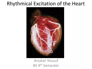
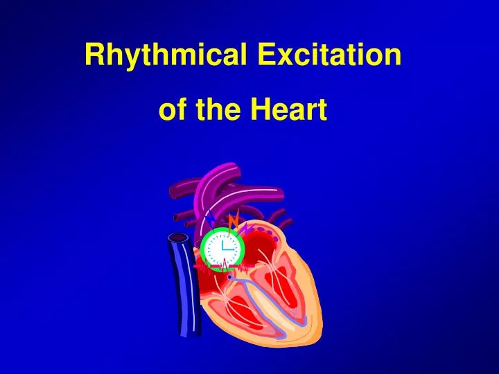
Rhythmical Excitation of the Heart. Specialized Excitatory and Conductive System of the Heart. Sinus node (Sinoatrial or S-A node) Internodal pathways between Sinus and A-V nodes Atrioventricular (A-V) node A-V bundle The left and right bundle branches Purkinje fibers.

E N D
Specialized Excitatory and Conductive System of the Heart • Sinus node (Sinoatrial or S-A node) • Internodal pathways between Sinus and A-V nodes • Atrioventricular (A-V) node • A-V bundle • The left and right bundle branches • Purkinje fibers
The Electrical System of the Heart Internodal Tracts SA Node Left Bundle Branch AV Node Anterior Superior Fascicle Bundle of HIS Posterior Inferior Fascicle Septal Depolarization Fibers Purkinjie Fibers Right Bundle Branch
Sinoatrial Node • Specialized cardiac muscle • Posterolateral wall of the right atrium • About 3 mm wide, 15 mm long and 1 mm thick • The fibers of this node have no contractile filaments • However, these fibers connect directly with the atrial muscle fibers • Automatic rhythmicity of the sinus fibers • Capability of self-excitation
Mechanism of Sinus Nodal Rhythmicity • Resting membrane potential of sinus nodal fibers between discharges has a negativity of -55 to -60 mV compared to -85 to -90 mV of the ventricular muscle • The cause of this negativity is leaky Na and Ca channels on the cell membrane • Fast Na channels • Slow Na-Ca channels • K channels • After action potential occurs, return of the potential to its negative state occurs slowly
Self-Excitation of Sinus Nodal Fibers • Na ions tend to leak inside • When the potential reaches a threshold voltage of about -40 mV, Na-Ca channels become activated • Why does this leakiness of Na-Ca channels not cause the sinus node to remain depolarized all the time? • Na-Ca channels become inactivated (they close) within 100-150 miliseconds • At the same time K channels open and movement of positive charges out of the cell causes “hyperpolarization” • This self-excitation cycle goes on and on throughout life
Signal Propagation in the myocardium from Lippincott ‘s Illustrated Reviews of Physiology
Internodal Pathways and Transmission of Cardiac Impulse Through the Atria • The ends of the S-A fibers connect directly with surrounding atrial muscle fibers • Velocity of conduction in most atrial muscle is about 0.3 m/sec • But it is 1 m/sec in several bands of the atrial fibers • Anterior interatrial band, passes to the left atrium • Three other small bands terminate in the A-V node • Anterior, middle and posterior internodal pathways
A-V node and delay of Impulse Conduction from the Atria to the Ventricles • Importance of the delay in cardiac impulse from the atrium to the ventricles • A-V node is located in the immediately behind the tricuspid valve (posterior wall of the right atrium) • The total delay in the A-V nodal and A-V bundle system is about 0.13 sec • In addition to the initial conduction delay of 0.03 sec (=0.16 sec) • Cause of the slow conduction: diminished numbers of gap junctions between successive cells in the conducting pathways
A-V node and delay of Impulse Conduction from the Atria to the Ventricles
A-V node and delay of Impulse Conduction from the Atria to the Ventricles
Rapid Transmission in the Ventricular Purkinje System • Special purkinje fibers • They are very large and can transmit 1.5 to 4 m/sec (150 times faster tha the A-V nodal fibers) • This rapid transmission of APs in Purkinje fibers is caused by high level of permeability of the gap junctions at the intercalated discs • The Purkinje fibers have very few myofibrils (and contract little or not during the course of impulse transmission) • One-way conduction through the A-V bundle • Role of continuous fibrous barrier
Distribution of the Purkinje Fibers in the Ventricles • The right and left bundle branches lie beneath the endocardium on the two respective sides of the ventricular septum • These branches in turn sidewise toward the apex of the heart • The elapsed time is about 0.03 second (from the entrance of cardiac impulse in the ventricular septum to terminations of the Purkinje fibers)
Transmission of the Cardiac Muscle in the Ventricular Muscle • Velocity of transmission is about 0.3 to 0.5 m/sec in the ventricular muscle fibers • Route of transmission from the endocardium towards epicardium • The total time for transmission of the cardiac impulse from the initial bundle branches to the last of the ventricular muscle fibers in the normal heart is about 0.06 second.
Control of Excitation and Conduction in the Heart • The Sinus node as the pacemaker of the heart: • A-V nodal fibers, when not stimulated from some outside source, discharge at an intricsic rhythmical rate of 40 to 60 times / min • The Purkinje fibers discharge at a rate of 15-40 times/min • Normal Sinus rate: 70-80 times/min • Faster self-excitatory nature of the S-A node • S-A node discharges again before either the A-V node or the Purkinje fibers can reach their own threshold of self-excitation
Abnormal Pacemakers – “Ectopic” pacemaker • Occasionally some parts of the heart develops a rhythmical discharge rate that is more rapid than that of the sinus node • In such cases, the pacemaker of the heart shifts from the sinus node to the A-V node or to the excited Purkinje fibers (Ectopic pacemaker) • Another cause of the shift of the pacemaker is the blockage of transmission of the cardiac impulse from the sinus node to the other parts of the heart • The new pacemaker occurs at the A-V node or A-V bundle
Stokes-Adams Syndrome • A-V block: Cardiac impulse fails to pass from the atrium to the ventricles • A new pacemaker usually develops in the Purkinje system of the ventricles (15-40 heart beats / min) • After a sudden A-V bundle block, a suppressed state occurs for 5 to 20 sec • Delayed pickup of the heartbeat is called Stokes-Adams syndrome
Synchronous Contraction of the Ventricular Muscle • Time period between excitation of the first and last ventricular muscles is 0.03 to 0.06 sec. • This causes all portions of the ventricular muscle in both ventricles to contracting at almost the same time • Effective pumping by the two ventricular chambers requires this synchronous type of contraction
Mechanism of Vagal Effects • Acetylcholine released at the vagal nerve endings greatly increases the permeability of the fiber membranes to potassium ions • This rapid leakage of K out of the conductive fibers causes hyperpolarization • In the S-A node, vagal stimulus makes the resting membrane potential more negative (-65 to -75 mV) • If the vagal stimulation is strong enough, it is possible to stop entirely the rhythmical self-excitation of the sinus node.
Mechanism of the Sympathetic Effects • Sympathetic stimulation increases • the rate of sinus nodel discharge • the rate of impulse conduction • the force of contraction • Stimulation of the sympathetic nerves releases norepinephrine at the sympathetic nerve endings • Norepinephrine increases the permeability of the membrane to Na and Ca ions • This causes a more positive resting membrane potential