Download
1 / 19
200 likes | 322 Views
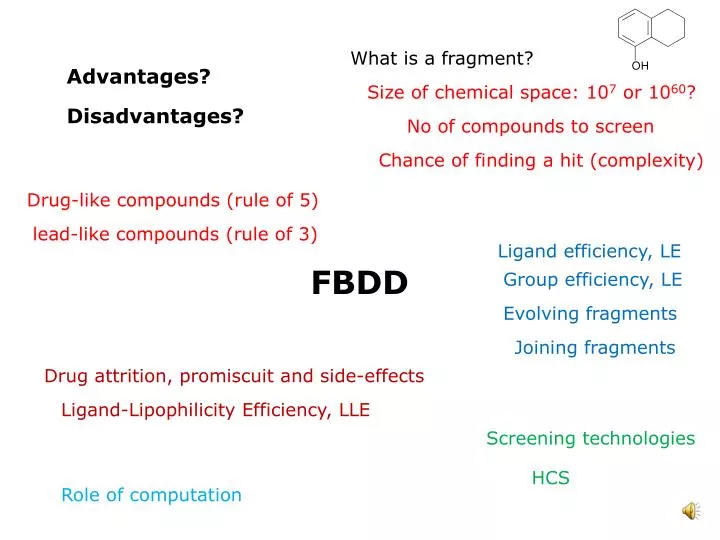
What is a fragment?. Advantages?. Size of chemical space: 10 7 or 10 60 ?. Disadvantages?. No of compounds to screen. Chance of finding a hit (complexity). Drug-like compounds (rule of 5). lead-like compounds (rule of 3). Ligand efficiency, LE. FBDD. Group efficiency, LE.

E N D
What is a fragment? Advantages? Size of chemical space: 107 or 1060? Disadvantages? No of compounds to screen Chance of finding a hit (complexity) Drug-like compounds (rule of 5) lead-like compounds (rule of 3) Ligand efficiency, LE FBDD Group efficiency, LE Evolving fragments Joining fragments Drug attrition, promiscuit and side-effects Ligand-Lipophilicity Efficiency, LLE Screening technologies HCS Role of computation
What is a fragment? (A) (B) Kd = 400 μM Kd = 2 mM • Fragments (e.g. (A), (B)) have: • low molecular mass (~100-200 Da) • typically low binding affinities (> 100 μM) (C) KI < 0.5 nM (C) Is a Bcl-XL inhibitor that has been in phase I/IIa cancer clinical trials; it was developed from fragments (A) and (B). This is a full-size drug-like/lead-like compound, not a fragment. (Aside: μM = 10-6 M; nM = 10-9 M; mM = 10-3 M)
Fragment – based drug design: 2 key ideas 1st key idea: The size of chemical space Lipinksi’s ‘rule’ of 5: a drug should have a Molecular mass less than about 500 g mol-1. How many isomers are there of C2H6 ___ C4H10 ___ C6H14 ___ C7H16?___ 1 2 4 7, including optical isomers How many potential drug-like molecules are there, i.e. with MM < 500 g mol-1 and therefore with less than ~30 heavy atoms (i.e. excluding H) ? i.e. how big is ‘chemical space’ for drug-like compounds? How many potential ‘fragments’ are there with < MM < 250 Da (g mol-1) ~12 heavy atoms? i.e. how big is chemical space for fragment-like compounds? 1060 107 It is feasible to screen a far greater proportion of fragment space compared to chemical space for drug-like compounds.
Fragment – based drug design: 2 key ideas 2nd key idea: Binding efficiency Because fragments are much smaller, they will bind to their target protein with lower affinity, typically μM – mM (rather than μM – nM for drug-like compounds that can form far more interactions). Consequently, the screening techniques employed in FBDD must be more sensitive than those used in a HTS assay. Nevertheless, the binding energy per atom can be as high as that for good drug-like hits. Generally, sensitive biophysical techniques are required (a) to detect weak binding and (b) to determine the binding interactions. X-ray crystallography and NMR are excellent for detecting low affinity binding of fragments; X-ray crystallography has the advantage that it also shows how the fragment bind. Surface plasmon reasonance (Biacore) (c.f. CAR’s BS133 lecture notes) is also used. Although good fragment hits bind weakly, they can still form good quality interactions with the target enzyme. Once weakly binding fragments have been identified, it is necessary to develop them into lead molecules, which will naturally be much more drug-like in size.
FBDD – motivation Success A number of biotech companies have used FBDD to develop clinical candidates Failures The optimism of HTS and combinatorial chemistry has not yielded the new drugs that had been expected in the 1990s – companies are looking for other approaches and FBDD is one of those actively being considered Disadvantages Difficult to screen low affinity compounds: biophysical approaches required. High investment in structural biology required to develop fragment hits into lead compounds as this requires understanding of molecular interactions and of the binding modes within the active site; also to eliminate false positives. Some targets are not amenable to 3D-structure determination. Advantages The need to screen only a small number of compounds means that small biotechnology companies and universities can do FBDD. (Universities are often good at biophysics).
HCS: high concentration screening HTS Can only detect compounds that bind with a reasonable affinity – and it will miss fragment hits that bind with a lower affinity. HCS The advantages of a smaller screening library can be partially obtained by using higher concentrations for the ligands in the screen – appropriate for ligands up to 350 Da. The use of higher concentrations will make it a little easier to detect the smaller hits that have lower affinity. This is a half-way house towards FBDD.
Lead-like compounds Lead-like compounds Most drug-like compounds have a molecular mass < 500 Da. But most lead compounds need to be modified to increase potency; usually involves adding functional groups and so increases the molecular mass. Starting a lead development programme with a compound of mass ~500 Da leaves no room to develop the compound (without first making it smaller). Starting with a mass of around 350 Da makes this development easier. Indeed, it has been observed that most lead-like hits have a molecular mass < 350 Da. (Note the ‘rule of 3’ for lead-like compounds: MM ≤ 300 Da, clogP ≤ 3, # H bond donors ≤ 3, # H bond acceptors ≤ 3, cf Lipinski rule of 5 for drug-like compounds) One strategy for following up a fragment hit is to find lead-like compounds (e.g. with mass ~ 350 Da) that are in some ways similar to the fragment hit – screening ~500 such compounds could be useful. (See the PC lab activities for ways of checking if compounds are similar; NB clogP is a programme to calculate log P)
Ligand efficiency, LE LE is a concept for comparing hits across different series of compounds and for assessing the effectiveness of compounds optimization. LE = -ΔG / HAC ≈ -RTln(IC50) / HAC Where HAC is the number of heavy atoms (or non-hydrogen atoms) ΔG is the free energy of binding of the ligand; ΔG will be –ve if the drug binds hence the –ve sign to make sure LE is a +ve number The units are kcal mol-1 (heavy atom)-1– the real world doesn’t always use SI units! and the literature often doesn’t give them, hence my omission below. For an oral drug with molecular mass < 500 Da (to fulfil Lipinski’s rules) and IC50 < 10 nM, LE should be at least 0.3. As the compound gets bigger during optimization, the idea is to ensure that LE does not decrease too much but rather stays above ~0.3.
Calculating Ligand efficiency, LE Using LE = -RTln(IC50) / HAC IC50 = 135 μM LE = - 8.314 * 298 * -8.910 / (1000 *4.184* 11) = 0.48 ln(135 10-6) = -8.910 (this is log to base e) (IC50 = 135 μM = 135 10-6 M) R = 8.314, T = 298, HAC = 11, by 1000 to covert from J to kJ and then by 4.184 to covert from kJ to kcal
Group efficiency, GE 0.28 1.6 0.54 0.42 0.32 1.5 GE = -ΔG / HAC, whereΔG is the contribution of a given group GE allows the estimation of an individual groups contribution towards the overall free energy of binding. (A) (B) IC50 = 135 μM IC50 = 80 μM The group efficiency of the methyl group, 0.32, was determined from the binding energy of the two fragments (A) and (B) that differ only in a methyl group at this position. • Assumptions: • that the total binding free energy of the whole ligand is the sum of the binding free energy of the groups – this will not always be true. • That the fragments (A) and (B) bind to the target in the same way – this will not always be true (but can be checked by X-ray crystallography).
Ligand-Lipophilicity Efficiency, LLE A significant part of protein-ligand binding involves desolvation of the ligand. Therefore, in general, provided that the shape of the drug is favourable, the more lipophilic a ligand is, the more favourably it will bind. A consequence of this is that lipophilic ligands could bind to any hydrophobic binding pocket; this could be in the target enzyme or conceivably in other enzymes. Thus, more hydrophobic drugs tend to be more promiscuous (i.e. bind to targets that they should not bind to. Analysis has shown that more lipophilic compounds carry a higher risk of drug failure as this is associated with nonspecific toxicity. Ligand-lipophicity efficiency is defined as LLE = pIC50 – clog P (or variants such as LLE = pKi – clog P or LLE = pKi – log D) where clogP is the log P calculated using the clogP programme
Using Ligand-Lipophilicity Efficiency, LLE Inappropriate physico-chemical properties are thought to be the main reason for attrition (compound failure) in drug development. LLE can be used to monitor a ligand during development to make sure that it does not become too hydrophobic. The target value for LLE is around 5-7 or greater. Problems with hydrophobicity can be avoided by choosing fragments that are not too hydrophobic. The mean clog P for recent drugs from large pharma is ~ 3.5 – 4.2. The mean clog P for patented compounds from Astex, a company based on FBDD was recently 2.4. This suggests that drugs derived from FBDD are likely to have fewer failures in development (or post development) and are likely to have fewer side effects. This may be an additional advantage of FBDD.
Complexity • The more complex a ligand is, the less likely it is to bind to a the target enzyme because • More chance of steric clashes • More chance of hydrogen bond mismatch • Large ligands will inevitably be complex. This reduces the chance of finding hits. • Fragments do not suffer from this complexity issue anywhere near as badly as lead-like ligands or drug-like ligands simply because they are so much smaller.
Optimization of fragments Evolving fragments Structural information, e.g. from X-ray crystallography is used to design larger molecules that make additional interactions. A requirement is that the fragment ‘anchor’ does not change binding mode during the fragment evolution. (This can be checked by X-ray crystallography). An example is the evolution of the β-secretase lead molecule (C) from (A) via (B). Note that LE should be monitored during fragment optimization to ensure that it stays > 0.3. (A) (B) (C) ~1 mM IC50 = 130 μM IC50 = 0.08 μM
Optimization of fragments Combining fragments Sometimes different fragments are found to bind to different parts of the target enzyme. They can be linked using a suitable linker. The difficulty is finding a suitable linker that allows both fragments to bind in their original positions without too much strain in the linker; the linker also needs to interaction favourably with the enzyme. An example is fragments (A) and (B) linked and then optimized to (D) before being developed into (C), a Bcl-XL inhibitor in clinical trial. (D) Kd = 6.9 μM (C) KI < 0.5 nM (A) (B) Kd = 400 μM Kd = 2 mM
Optimization of fragments Fragment tethering Uses the formation of a disulfide bond between a chemically reactive fragment and a cysteine residue in the target protein. Fragments with the greatest affinity for the protein within the vicinity of the cysteine form stable disulfide bonds. These are detected by mass Spectrometry. Fragments react with (A) bound to enzyme, e.g. B) (A) – reacts with enzyme, Enz (B) Finally, (D) from (C) IC50=0.005 μM (C) Developed from (B) IC50 = 0.19 μM (D) caspase-1 inhibitor
Using fragments to identify new binding sites Heat Shock Protein 90 (HSP 90) inhibitors (A) Was discovered using an NMR-based screening approach A fragment library was screened with (A) already bound – (B) was found to bind tightly in presence of (A) but weakly in absence of (A), showing cooperative effects. (C) Was designed using structural information from NMR and X-ray to effectively join the two fragments – but note some improvements to the part that came from (B). (A) Kd = 20 μM (B) Kd = 150 μM (with A) Kd > 5000 μM (without A) (C) Kd = 4 μM
Computational approaches • Used to design fragment libraries, e.g. by analysing existing drugs and decomposing them into constituent fragments • Docking (virtual screening) to • identify possible fragments that might bind • (b) Suggest the binding mode of fragments known to bind to generate ideas on how to grow the fragment • (c) To check the predicted binding mode of compounds suggested by analysis of the X-ray structure. • Docking programs were developed to work on drug-like and lead-like compounds so do not always work on fragments
Screening technologies Surface plasmon resonance For methodology see BS133_CAR_audio_SPR_4A.ppt* Useful when there is no structure. Requires smaller amounts of protein than X-ray crystallography or NMR. Can determine enthalpy and entropy of binding. Can also determine kinetics of binding – e.g. this will show if there is a slow off-rate for the ligand. NMR The NMR signal of a ligand can be monitored – it will change if the ligand is displaced by a fragment. E.g. in HSP 90 it is possible for fragments to displace ADP which binds with low affinity – changes in the ADP NMR signal indicate that a fragment has bound. Why is it important that ADP binds weakly? X-ray-crystallography Expensive but essential for confirming the fragment binding mode and for generating ideas for fragment optimization. Generally carried out by soaking the compounds into crystals. The fragment binds weakly so cannot displace a ligand that binds tightly