Download
1 / 25
250 likes | 411 Views
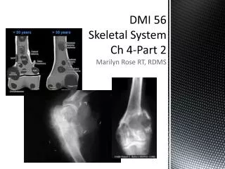
Ch. 4-DMI 56 Skeletal System. Marilyn Rose RT, RDMS. Physiology of Skeletal System Congenital/ Hereditary Diseases of Bone Inflammatory and Infectious Disorders Metabolic Bone Disease Lead Poisoning. Outline- Part 1.

E N D
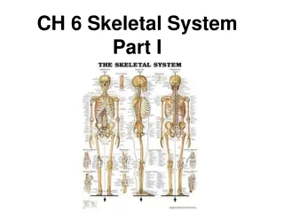
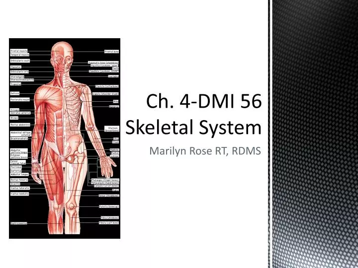
Ch. 4-DMI 56 Skeletal System Marilyn Rose RT, RDMS
Physiology of Skeletal System • Congenital/ Hereditary Diseases of Bone • Inflammatory and Infectious Disorders • Metabolic Bone Disease • Lead Poisoning Outline- Part 1
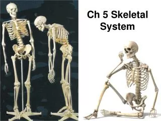
Composed of two highly specialized connective tissues- Bone and cartilage • Bone- organic matrix + inorganic salts- Ca++ and phosphate • Perisoteum with blood vessels- covers outer surface- except joints • Shaft= diaphysis= medullary cavity (marrow) • Ends= ephipysis • Compact bone= outer layer- haversian systems • Spongy bone= inner weblike marrow space with thin trabaculae Physiology of Skeletal System
Bones form from hyaline cartilage • Primary ossification @ 8th week of life is the center of cartilage • Entire shaft is ossified at birth • Secondary center is at epiphyses • Remains separate from diaphysis by epiphyseal cartilage until bone stops growing- then epiphysis an diaphysis fuse- where they meet is the metaphysis. • Bones grow in diameter by • osteoblasts- form new bone - osification • osteoclasts- enlarge cavity- resorption • Bone age is determined by the atlas Greulich and Pyle • After 40 years- bone loss > bone gain Physiology contd.
Vertebral anomalies • Spina bifida • Osteopetrosis • Osteogenesisimperfecta • Achondroplasia • Congenital Hip Dysplasia Congenital / Hereditary Disease of Bone
Transitional vertebrae • Has characteristics of both sides of a major division of the spine • Occurs @ • Lumbosacral junction- expanded transverse process- form unilat or bilat joints with the sacrum. • C-spine -7th cervical vertebrae- rudimentary rib • Both are incidental findings • Lumbosacral - degenerative changes in the hip • Cervical can compress the brachial nerve plexus or subclavian artery…< blood flow…Thoracic outlet syndrome Vertebral anomalies
Posterior defect of spinal canal • Failure of posterior elements to fuse • oculta- mild= splitting bony canal at L5-S1 • Meningocele- herniation of meninges • Myelomeningocele- herniation of meninges and portion of the spinal nerve roots. • Dimpling or tuft of hair will often be present at the site of the spinal defect. • Associated with.. club foot, gait disturbances and bladder/ bowel incontinence (neurogenic) • Myelomeningocele has neurologic deficits and almost always associated with Chiari II malformation- caudal displacement of posterior fossa. • Hydrocephalus is a common complication Spina Bifida
Marble Bone • Rare hereditary bone dysplasia • Failure of resorptive mechanism of calcified cartilage interferes w/ normal replacement by mature bone. • Bones are brittle/ stress fractures • Anemic • Range in severity and age of clinical presentation- fulminant/ fatal – incidental finding. • Radiographic- symmetric generalized increase in bone density • > mA and kVp Osteopetrosis
Brittle bones • Inherited- generalized disorder of connective tissue • Characterized by multiple fractures and unusual blue color to the sclera of the eye. • Adults are in a wheelchair- skeleton does not support the body weight • Radiographic- repeated fx, severe osteoporosis due to thin defective cortex • Fx heal with callus- causing bizarre deformities • < kVp to compensate for bone loss • Wide skull sutures • Can be confused with child abuse OsteogenesisImperfecta
Most common form of dwarfism • Diminished proliferation of cartilage in the growth plate (< enchondral bone formation) • Autosomal dominant • Does not affect membranous bone formation- short limbs with a normal size trunk (axial skeleton) • Large head w/ frontal buldging, saddle nose, prognathous (jutting jaw), prominent jaw • Radiographic-Erlenmeyer flask deformity, spondylosis Achondroplasia
Incomplete acetabulum formation • caused by • physiologic - > hormone at delivery • mechanical – low amniotic fluid or breech • After delivery the hips are mechanically rotated and a pop or hip click can be heard • The tendons and ligaments are affected • Ultrasound is the best method at one month of age • If hip dysplasia is diagnosed- a Velcro Pavlik harness is used to realign the joint space Congenital Hip Dysplasia 3 4 2 1
Rheumatoid Arthritis • RA variants- • Ankylosing Spondylitis • Reiter’s Syndrome • Psoriatic Arthritis • Osteoarthritis (DJD) • Infectious Arthritis • Bursitis • Rotator cuff • Menisci tear • Bacterial Osteomyelitis • Tuberculous Osteomyelitis Inflammatory and Infectious Disorders
Chronic systemic disease of unknown cause • Appears as an noninfectious inflammatory (synovial membranes) arthritis of the small joints of the hands and feet • 3X more often in females • Onset is in 40’s • May be very progressive or can undergo spontaneous remissions of variable lengths • Symmetric involvement of multiple joints • Progressing towards the trunk • Synovitis- --excessive exudate---inflamm----synovium to >--- mass of thick tissue cause erosion of articular cartilage- erosion from lytic enzymes Rheumatoid Arthritis
Ankylosing Spondylitis • Almost always starts in the SI joints • Bilat / symmetric involvement • Narrow joint space- progress to L spine • Ossification in paravertebral tissue / lateral bony bridges/ syndesmophytes- produce the “bamboo spine” • Reiter’s Syndrome • Reactive arthritis • Arthritis, urethritis and conjunctivitis • Affects young adult males- post venereal or GI infections • SI joints, heel, toes • Unilateral SI involvement w/o spine changes • Psoriatic Arthritis • RA with skin changes of psoriasis • Distal interphalangeal joints of hands and feet • Erosion and hypertrophic changes at tendon/ lig insertion RA Variants
Extremely common • Loss of joint cartilage and reactive new bone formation • Wear and tear of aging • Weight-bearing joints- spine, hip, knee and ankle…interphalangeal joints of the fingers • Repeated trauma • Abnormal stress • Result of septic or inflamm arthritis • Radiographic: narrow joint space, bony spurs (osteophytes) • Erosions can cause cyst-like lesions Osteoarthritis (DJD)
Pyogenic orgainisms • Hematogenous route or adjacent osteomyelitis or post surgery or trauma of joint • Abrupt, high fever • Tender, swollen, joint (s) • Most common type is migratory arthritis from Lyme disease • Radiographic- soft tissue swelling • In children- fluid distention of joint capsule • 8-10 days after onset- focal erosion in cortex • Severe, untreated- extensive destruction and loss of cortical outline • Tuberculous arthritis: • Chronic, gradual onset- one joint- spine, hip or knee • Juxtaarticular osteoporosis precedes bone destruction • Joint effussion • Disorganization of join Infectious Arthritis
Inflammation of bursae • Small fluid filled sacs near joints • Reduce friction • Cause: • Repeat physical activity • Trauma, RA, gout or infection • Tenosynovitis not seen on plain xray • Ultrasound is BEST • Radiographic: deposits of Ca++ in tendons, frozen joint • Early stages shows bursae filled with fluid Bursitis
Shoulder- muscular, tendenous • Muscles: • Teres minor, infraspinatus, supraspinatus, subscapularis • Rupture = communication btw shoulder joint and subacromialbursae - demonstrated by orthrography- Inject contrast into joint space • MRI is the modality of choice • Tear= high signal intensity • US is preferred initially to see tear of tendon • Knee pain • Acute trauma, more frequently degenerative • Inherent in human knee function • MRI modality of choice (90-98% accuracy) • Tear sharp line of high signal intensity • Crosses normally dark triangular meniscus • Show ACL/ PCL and changes in the underlying bone • US may demonstrate tenosynovitis- thick fluid filled Rotator Cuff/ Menisci Tears
Bacterial • Inflammation of bone and bone marrow • Hematogenous- adjacent infection/ direct intro of organisms (OR) • Acute – rich red marrow • Infants- metaphysis- femur/ tibia- staf/ strep • Fever/ tender • Adult- vertebrae- localized back pain and back spasm- rarely long bones • Decreased with antibiotics • Complication of IV drug use • Diabetic- soft tissue infection spread from skin abscess- cellulitis- osteomyelitis • Radiographic- deep soft tissue swelling adjacent to metaphysis, displacement of fat planes • Tuberculous • Very rare today • Thoracic or lumbar spine • Pott’sdz- TB of spine- irregular, bone destruction • Eventual vertebral collapse Bacterial/ Tuberculous Osteomyelitis
Osteoporosis • Osteomalacia • Rickets • Gout • Paget’s Metabolic Bone Disease
Generalized, localized deficiency of bone matrix • Mass of bone per unit volume is decreased • Accelerated resorption of bone- • decreased bone formation • Cushings syndrome • Prolonged steroid • Disuse or immobilization (cast) • Loss of mineral salts- more lucent • 50-70% lost before lucent areas are seen on radiographs • Use LOWEST kVp – short scale to look for lucency • DEXA is often used- measure bone mineral content • Radiographic- cortical thinning- spine/pelvis- anterior wedging or compression fractures Osteoporosis
Insufficient mineralization adult skeleton • Failure of calcium/ phosphorus deposition • From inadequate intake • Failure of absorption • Vitamin D necessary for intestinal absorption • Chronic renal failure • Renal cause for calcium lost in urine • Radiographic- loss of bone density due to nonmineralized osteoid • Thin but prominent cortex • Bones bend and display bowing deformity • Rickets is systemic disease of infancy / childhood that is equivalent to osteomalacia • Lack of UV rays for vitamin D • Radiographic- increase in distance between ossified epiphysis and end of shaft • Bowing of weight bearing bones Osteomalacia/ Rickets
Disorder in metabolism of purine • Increase in blood level of uric acid • Deposition of uric acid crystals in joints, cartilage and kidney • Causes of hyperuricemia • Metastatic carcinoma • Myeloma • Hemolytic anemia • Drugs- chemo/ HTN • Kidney failure • Radiographic- painful arthritis • Single joint- first metatarsophalangeal joint • “rat bite” erosions • Large soft tissue swelling- tophi Gout
Osteitisdeformans • Common chronic metabolic disease of the skeleton • Destruction of bone • Repair • Weak, deformed, thick structures that fracture easily • 2x in men • Pelvis and weight bearing bones • Radionuclide bone scan is most efficient method to see extent of lesions • Radiographic- mottled, cotton- wool appearance- cold (destructive) hot (repairative) • Increased trabeculation • Picture frame vertebrae Paget’s
Especially lead paint • Occupational inhalation of fumes • Drinking water with lead pipes • Eating food prepared, processed, stored in lead containers • Number one environmental pollutant worldwide • Chronic- mental retardation, seizures, behavioral disorders or delayed development • Radiographic- in children lead is deposited in growing portions of skeleton- metaphyses- lead lines • Pica kids may eat the paint and find abdominal opacities like barium (CT) Lead Poisoning