Download
1 / 12
120 likes | 431 Views
Part II. Monitoring and Optimizing Detergent Concentration While Following Protein Homogeneity.

E N D
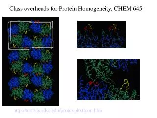
Part II. Monitoring and Optimizing Detergent Concentration While Following Protein Homogeneity Larry Miercke*, Rebecca Robbins*, Cory Anderson, Akanksha Bapna1, Vladimir Denic2, Robert Edwards1, Bill Harries, Frank Hays, Joe Ho, Mimi Ho, Min Li3, Lynn Martin, Zach Newby4, Joseph O’Connell, Edwin Rodriquez, Andrew Sandstrom5, Meseret Tessema, Tomomi Tsomeya, Yaneth Robles, Dave Savage, Ronald Yeh and Robert Stroud Department of Biochemistry and Biophysics, 1Department of Physiology, University of California San Francisco, San Francisco, CA; Current addresses are 2 Molecular and Cellular Biology, Harvard University, Cambridge, MA; 3Novartis Vaccines and Diagnostics, Emeryville, CA; 4Novartis Institute for Biomedical Research, Cambridge, MA; 5Biochemistry & Molecular Biology, University of Chicago, Chicago, IL *Performed TDA data acquisition and analysis This work was supported by NIH Roadmap grant P50 GM073210 Please e-mail Larry at merc@msg.ucsf.edu for questions, comments ect.
Abstract Detergent concentration is critical to growing quality membrane protein crystals. Since the optimal detergent concentration lies just below the detergent phase boundary (Berger et al., 2006, Protein Science 15, 2682-2696), the starting detergent concentration must be minimized for initial crystallization trials. However, the majority of Pure Homogenous Stable targets contain excessive levels of detergent micelles upon concentration using molecular weight cut-off filters. Size Exclusion Chromatographyand a Tetra Detector Array/Analysis (refractometer, viscometer, light scattering, and UV detectors) are now being successfully utilized to monitor and optimize detergent concentration while assaying PDC homogeneity during purification and concentration for crystallization. In doing so, the oligomeric state, size, shape and the detergent:protein ratio of the Protein Detergent Complex is measured. Five different membrane proteins using 3 different detergents (OG, DDM, and FC14) and 4 different methods will be presented where detergent was successfully minimized while maintaining PDC homogeneity. Methods utilized where ultra filtration (centrifugal and high-pressure MWCO filters) plus SEC and dialysis, changing detergent isomer, Ni-NTA and ion exchange chromatography. Detergent micelle SEC retention volume, dn/dc, Rh, IV, mass and behavior on different ultrafiltration molecular cut-off filters and formats are all being measured using TDA. As expected, there is a direct correlation of measured excess micelle concentration to crystal phase separation and diffraction quality. An important discovery is that free micelles in the presence of PDCs tend to be highly retained on MWt cut-off filters which would freely pass a pure detergent micelle system; therefore, when measuring whether a micelle is retained or passed through by specific molecular cut-off filters and formats, it must be measured using a PDC system and not just buffered detergent controls.
Figure 1. TDAnalysis of OG. All TDAgrams acquired on SDX 200 using 20mM Hepes7.3, 150mM NaCl, 40mM OG solvent and 120 mM stock OG (23.39 mg/ml). A) TDAgram of 1.75 mg (75 μl stock) OG. B) Overlay of the RI traces of 5 different OG injections used to calculate dn/dc (inset; dn/dc = 0.157 ml/g; y intercept = 0.596; RSQ = 0.999). Note that a 40 mM OG injection gave no signal in all four detectors. C). Tabulated “whole peak” analysis of the biophysical parameters of an OG micelle, with errors less ≤ 1%. The micelle molecular weight (MMW) decreased with a decrease in concentration as expected (previous DLS & TDA studies; Lorber, Bishop & DeLucus,1990, BBA 1023, 254-65). B A RI area (mVml) OG (g) RALS RI 280nm DP C
Table 1. Detergent micelle retention volume of common detergent/column/buffer systems. This can be used to approximate the RV of your micelle (+/- lipids) SEC system.
Figure 2. TDA profile and analysis of ahuman 7-crosser expressed in Pichia and purified in OG. This is a prime example showing the effect of using different ultrafiltration formats and membrane types, and the effect on concentrating micelles in the presence of a PDC. Since the excess micelle peak is well separated from the PDC, the PDC can be concentrated prior to SEC, and detergent may be minimized by SEC. Ultimately, the detergent in the purified truncated PDC was minimized by concentrating prior to SEC using a 50kDa MWCO filter, further concentrated by 50kD post SEC, and over night 50kDa dialysis. This yielded an PHS sample with minimized detergent (10mM) that showed no detergent phase separation, and produced a 1.8 Å structure (Ho et al, submitted). Figure 2 caption continued on next slide
Figure 2 cont. A) Superdex TDAgram in 40mM OG post 40x 50kDa stir concentrate shows the PDC to be a homogenous (single Gaussian 280nm peak) and globular (IV=0.05) tetramer with a 5.7 nm Rh; it binds 260 moles OG, has a 0.17 ml/g dn/dc, and contains 38mM excess OG micelles. B) Flowchart diagram shows the 3x difference in the final OG micelle concentration between using a 50kDa XM stir and YM spin ultrafiltration membrane to concentrate the PDC, the excessive micelle concentration upon the final 10x concentration factor, and the stark effects of excess micelles (270mM verses 67mM verses 10mM) on crystallization. C0 is the non-concentrated pooled fractions post SEC, Ci is the intermediate concentration and Cf is the final concentrated sample which enters the crystallization trials. Note that pure OG micelles do not concentrate on a 50kDa, but do concentrate in the presence of a PDC.
3.4 Å Figure 3. An example where the detergent was successfully minimized using a different detergent isomer. The TDAgrams profile a 12 crosser post 100kDa spin concentration in 0.5mM β-DDM and α-DDM using a 0.75x60cm TSK G3000SW column. In β-DDM (A), the PDC is homogenous (peak 1 with 280nm signal), β-DDM micelles concentrate on 100kDa (peak 2 with large RI and DP signal), extreme phase separation is exhibited during crystallization and no crystals are grown. In the absence of protein, β-DDM micelles are not retained. When purified in α-DDM using identical procedures (B), α-DDM micelles do not concentrate on a 100kDa filter (peak 3; no 280nm and minimal RI and RALS), the PDC is mostly homogenous (peak 2) with a small void (peak 1, minimal 280nm and maximum light scattering signal), no phase separation is observed during crystallization and 3.4 Å crystals are grown.
Figure 4. An example using TDA and cation exchange to successfully minimize DDM detergent concentration while concentrating protein and maintaining homogeneity. Continued on next slide.
Figure 4 continued. This is a prime example showing that the detergent concentration may need to be increased when concentrating protein chromatographically. The protein is an eukaryotic 12-crosser over-expressed in S. cerevisiae. All TDA profiles were obtained using a Superdex 200 10/300 GL column in 2mM β-DDM. A) TDAgram after being concentrated 58x post SEC using a 50kDa XM filter and stirred cell. The pure PDC (inset) remains homogenous (Gaussian 280nm peak). The PDC sample contains 61mM excess DDM micelles, which represent 47% theoretical removal; only 25% was removed using an YM 50kDa spin filter (TDAgram not shown). It produced major detergent phase separation and yielded no crystals. B) TDAgram after being concentrated using S cation exchange (inset) and 2mM β-DDM buffer. The PDC eluted from S at 9 mg/ml (inset) and contains -1.5 mM DDM (negative RI peak). Due to a low detergent: protein ratio the PDC forms highermers. No detergent phase separation or crystals were formed upon crystallization trials. C) TDAgram after being concentrated to 7.5 mg/ml using S cation exchange and 4mM β-DDM buffer. The PDC remains homogenous and contains no excess detergent micelles. No crystals or detergent phase separation were produced from the first round of crystallizations; however, this project is now worthy to enter comprehensive crystallization trials. D) Table of TD whole peak analysis (verses peak slice) using 7 independent preparations and 8 TDAgrams. Errors for the five measured parameters range from 5.0 to 11.0 %. For analysis, the protein component was assumed to have a MWt, dA/dc and dn/dc of 43.6kDa, 1.30 OD280nm/mg and 0.185 ml/g, respectively. Measured DDM dn/dc values , which were dependent on running conditions, ranged from 0.133 to 0.157 ml/g and the micelle MWt ranged from 80-90kDa (157-176 monomers/micelle). As expected for a 85kDa β-DDM micelle, DDM is not dialyzable using a 50kDa bag: after 9 day PDC dialysis the DDM micelle concentration was reduced by only 8% (168mM verses 155mM).
Figure 5. TDAgrams of a different eukaryotic 12 crosser in 0.5mM FC14. A) Superdex TDAgram post 54x stirred 50kDa concentration and 4 day dialysis. The functional PDC isamostlyhomogenous and slightly elongated dimer (IV=0.07) with 212 moles C14PC, and contains 9 mM excess FC14 micelles. Even though pure FC14 micelles (46-51kDa measured MMW) do not concentrate, 42 % of the micelles are retained upon concentration in the presence of a PDC. This sample produced major phase separation during crystallization trials and no crystals. B) TDAgram at 5mg/ml post concentration by affinity chromatography and desalt. The dimeric PDC remains >95% homogenous with excess micelles slightly below (negative RI peak) the mobile phase concentration of 0.5mM FC14. This target is now worthy for comprehensive crystallization trials.
Figure 6. 3TM/40mM OG TDAgrams. Continued on next slide. A. B. LALS RALS LALS RI 280nm PDC RALS DP Excess micelles OG micelle RALS RI C. D. 280nm OG micelle PDC DP RI 280nm PDC multimers DP
Figure 6. An example using TDA and anion exchange to successfully minimize OG detergent concentration while concentrating protein and maintaining homogeneity. The MP target was initially expressed in cell free, and then in E. coli for production. All TDA profiles were obtained using a Superdex 200 10/300 GL column in 40mM OG. A) TDAgram after 32x concentration on a 50kDa spin filter; the PDC remains homogenous as seen by a Gaussian UV trace (shown in purple) and contains 150mM excess OG. B) TDAgram after 2 day, 50kDa dialysis; only 60mM of the 150mM excess OG micelles was removed. The sample produced major phase separation during crystallization trials and produced no crystals. C) TDAgram following concentration to 11mg/ml using S/pH 5; the sample contains -13mM OG (or 27mM) and is composed of at least 3 PDC species. The SEC profile is not reversible with the addition of 40 or 150mM OG. D) TDAgramfollowingQ (pH 7.3, 55mM OG) at 16.7 mg/ml; the PDC is a homogenous and globular (IV=0.05) tetramer with a 4.3 nm Rh; the PDC binds 113 moles OG, has a 0.17 dn/dc and contains 8mM excess OG micelles.