Download
1 / 1
10 likes | 112 Views
Photoblinking / Photobleaching mathematical model for fluorescence images of microscopy. Isabel Rodrigues and J.Miguel Sanches Institute for Systems and Robotics Department of Bioengineering, Instituto Superior Técnico, Technical University of Lisbon Portugal. Abstract

E N D
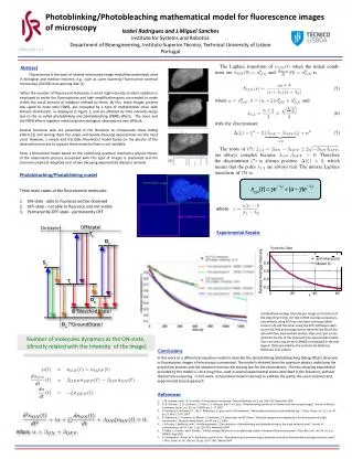
Photoblinking/Photobleaching mathematical model for fluorescence images of microscopy Isabel Rodrigues and J.Miguel Sanches Institute for Systems and Robotics Department of Bioengineering, Instituto Superior Técnico, Technical University of Lisbon Portugal Abstract Fluorescence is the basis of several microscope image modalities extensively used in biological and medical research, e.g., such as Laser Scanning Fluorescence confocal microscopy (LSFCM) and spinning disk [1]. When the number of fluorescent molecules is small, high intensity incident radiation is employed to excite the fluorosphores and high amplification gains are needed to make visible the small amount of radiation emitted by them. By this, these images present low signal to noise ratio (SNR), are corrupted by a type of multiplicative noise with Poisson distribution, as displayed in Figure 1, and are affected by time intensity decay due to the so called photoblinking and photobleaching (PBPB) effects. The noise and the PBPB effects together make long time biological observations very difficult. Several functions laws are presented in the literature to compensate these fading effects [2] and among them the single and double decaying exponentials are the most used. However, a simple and tractable theoretical model based on the physics of the observation process to support these empirical laws is not available. Here, a theoretical model based on the underlying quantum mechanics physics theory of the observation process associated with this type of images is presented and the common empirical weighted sum of two decaying exponentials (DExp) is derived. Photobleaching/Photoblinkingmodel Three main states of the fluorescence molecules: ON-state - able to fluoresce and be observed OFF-state - not able to fluoresce and not visible Permanently-OFF-state - permanently OFF. where Experimental Results Synthetic Data Standardized average intensity per image as a function of the experiment time, for two LSFCM real data sequences, one without using FLIP nor any other technique (dark circles in A) and the other using the FLIP technique (dark stars in B). Red and orange curves stand for the fits of the data with two exponentials models. Blue and cyan curves stand for the fits of the data with one-exponential models. The root mean square error (RMSE) is displayed in the plot legend. (Data provided by the Instituto de Medicina Molecular (UL) Lisbon). Number of molecules dynamics at the ON-state,(directly related with the Intensity of the image) Conclusions In this work an a differential equation model to describe the photoblinking/photobleaching fading effects observed in fluorescence images of microscopy is presented. The model is derived form the quantum physics underlying the acquisition process and the obtained intensity decreasing law fits the observations. The two decaying exponential provided by the model is, since long time, used in several experimental works described in the literature, without theorectical reasoning . In this work, a theoretical model is derived to validate the justify the usual empriacl and experimental based approach References J. W. Lichtman and J.-A. Conchello, “Fluorescence microscopy,” Nature Methods, vol. 2, pp. 910–919, November 2005. N. B. Vicente, J. E. D. Zamboni, J. F. Adur, E. V. Paravani, and V. H. Casco, “Photobleaching correction in fluorescence microscopy images,” Journal of Physics: Conference Series, vol. 90, no. 012068, pp. 1 – 8, 2007. S. Gavrilyuk, S. Polyutov, P. C. Jha, Z. Rinkevicius, H. gren, and F. Gel’mukhanov, “Many-photon dynamics of photobleaching,” J. Phys. Chem., vol. 111, no. 47, pp. 11 961–11 975, 2007. X. Brokmann, J.-P. Hermier, G. Messin, P. Desbiolles, J.-P. Bouchaud, and M. Dahan, “Statistical aging and nonergodicity in the fluorescence of single nanocrystals,” Physical review letters, vol. 90, no. 12, 2003. J. Schuster, J. Brabandt, and C. von Borczyskowski, “Discrimination of photoblinkingand photobleaching on the single molecule level,” Journal of Luminescence, vol. 127, no. 1, pp. 224–229, November 2007. P. Didier, L. Guidoni, and F. Bardou, “Infinite average lifetime of an unstable bright state in the green fluorescent protein,” Phys. Rev. Lett., vol. 95, no. 9, p. 090602, Aug 2005. R. Zondervan, F. Kulzer, M. A. Kol’chenko, and M. Orrit, “Photobleaching of rhodamine 6g in poly(vinyl alcohol) at the ensemble and single-molecule levels,” J. Phys. Chem. A, vol. 108, no. 10, pp. 1657–1665, March 2004.


