Download
1 / 1

E N D
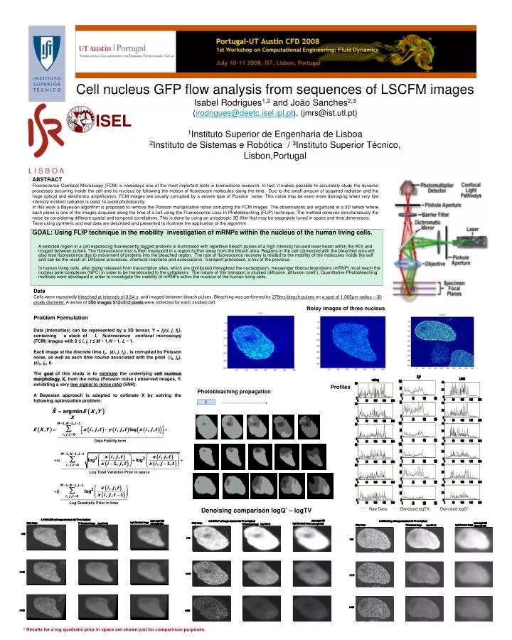
Raw Data, Denoised logTV, Denoised logQ* Cell nucleus GFP flow analysis from sequences of LSCFM imagesIsabel Rodrigues1,2 and João Sanches2,3(irodrigues@deetc.isel.ipl.pt), (jmrs@ist.utl.pt)1Instituto Superior de Engenharia de Lisboa2Instituto de Sistemas e Robótica / 3Instituto Superior Técnico, Lisbon,Portugal ABSTRACT Fluorescence Confocal Microscopy (FCM) is nowadays one of the most important tools in biomedicine research. In fact, it makes possible to accurately study the dynamic processes occurring inside the cell and its nucleus by following the motion of fluorescent molecules along the time. Due to the small amount of acquired radiation and the huge optical and electronics amplification, FCM images are usually corrupted by a severe type of Poisson noise. This noise may be even more damaging when very low intensity incident radiation is used to avoid phototoxicity. In this work a Bayesian algorithm is proposed to remove the Poisson multiplicative noise corrupting the FCM images. The observations are organized in a 3D tensor where each plane is one of the images acquired along the time of a cell using the Fluorescence Loss In Photobleaching (FLIP) technique. The method removes simultaneously the noise by considering different spatial and temporal correlations. This is done by using an anisotropic 3D filter that may be separately tuned in space and time dimensions. Tests using synthetic and real data are described and presented to illustrate the application of the algorithm. GOAL: Using FLIP technique in the mobility investigation of mRNPs within the nucleus of the human living cells. A selected region in a cell expressing fluorescently tagged proteins is illuminated with repetitive bleach pulses of a high intensity focused laser beam within the ROI and imaged between pulses. The fluorescence loss is then measured in a region further away from the bleach area. Regions in the cell connected with the bleached area will also lose fluorescence due to movement of proteins into the bleached region. The rate of fluorescence recovery is related to the mobility of the molecules inside the cell and can be the result of: Diffusion processes, chemical reactions and associations, transport processes, a mix of the previous.In human living cells, after being released from transcription sites, which are distributed throughout the nucleoplasm, messenger ribonucleoproteins (mRNP) must reach the nuclear pore complexes (NPC) in order to be translocated to the cytoplasm. The nature of this transport is studied (diffusion, diffusion coeff.). Quantitative Photobleaching methods were developed in order to investigate the mobility of mRNPs within the nucleus of the human living cells. DataCells were repeatedly bleached at intervals of 3.64 s and imaged between bleach pulses. Bleaching was performed by 279ms bleach pulses on a spot of 1.065μm radius – 30 pixels diameter. A series of 350 images 512×512 pixels were collected for each studied cell. Noisy images of three nucleus Problem Formulation Data (intensities) can be represented by a 3D tensor, Y = {y(i, j, t)}, containing a stack of L fluorescence confocal microscopy (FCM) images with 0 ≤ i, j, t ≤ M − 1,N − 1, L − 1. Each image at the discrete time t0, y(i, j, t0) , is corrupted by Poisson noise, as well as each time course associated with the pixel (i0, j0), y(i0, j0, t). The goal of this study is to estimate the underlying cell nucleus morphology, X, from the noisy (Poisson noise ) observed images, Y, exhibiting a very low signal to noise ratio(SNR). A Bayesian approach is adopted to estimate X by solving the following optimization problem: Profiles Photobleaching propagation t Denoising comparison logQ* – logTV * Results for a log quadratic prior in space are shown just for comparirson purposes.