Download
1 / 17
340 likes | 1.03k Views
Schistosomiasis. By, Cheryl Poleschuk Linda Hansen. Background. More than 200 million people are infected with schistosomiasis worldwide One of the WHO neglected diseases of the world Blood fluke = common name Also called, bilharzia 3 species cause schistosomiasis

E N D
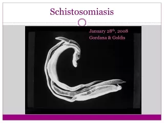
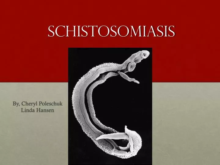
Schistosomiasis By, Cheryl Poleschuk Linda Hansen
Background • More than 200 million people are infected with schistosomiasis worldwide • One of the WHO neglected diseases of the world • Blood fluke = common name • Also called, bilharzia • 3 species cause schistosomiasis • S. mansoni- most wide spread • S. japonicum • S. haematobium
Geographical distribution • S. Mansoni • Africa • South America • Middle East • Caribbean • S. haematobium • Africa • Middle East • S. japonicum • South East Asia
S. haematobium S. japonicum
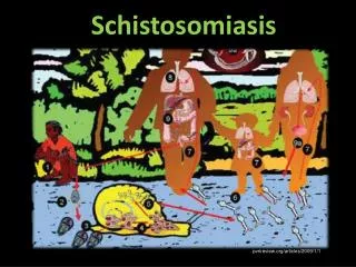
Hosts • Definitive- human • Intermediate- freshwater snails • Biomphalaria • Bulinus, Physopsis • Oncomelania • Vector- none • Reservoir • For S. japonicum- dogs, cats, rodents, pigs, horse, and goats
Source of Infection • Schistosome penetrates skin
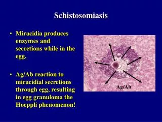
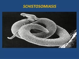
Morphology cont. • Adult female = 7-20mm long • Adult male = slightly shorter • Gynecophoral canal- ventral, longitudinal groove where the female normally resides • Eggs • S. mansoni has a lateral spine near the posterior end and are 114-180 microns long by 45-70 microns wide • S. japonicumare more round, harder to see spine, and are 70-100microns by 55-64 microns • S. haematobiumhas a terminal spine, 110-170 microns by 40-70 microns • Cercariae has a forked tail
Symptoms • Migratory phase- from time schistosoma penetrates until maturity and egg production • Days after infection • Rash or itchy skin • People are often symptomless • Acute phase (Katayama fever)- when schistosomes produce eggs around 4-10 weeks after initial infection • Chills • Fever • Fatigue • Headache • Malaise • Muscle aches • Lymphadenopathy • Gastointestinal discomfort
Symptoms cont.. • Chronic phase • Commonly asymptomatic • Intestinal schistosomiasis • Mild, chronic, bloody diarrhea • Mild abdominal pain • Lethargy • Schistosomiasishaematobia • Pain on urination • Hematuria (blood in urine) • Children repeatedly infected can develop anemia, malnutrition, and learning difficulties • About 8% are serious cases where the portal blood is impeded • Cirrosis, portal hypertension, enlargement of the spleen, ascites (accumulation of fluid in the abdominal cavity
Diagnosis • Microscopic identification of eggs in stool/urine • Concentration technique • Antibody detection • ELISA • Biopsy • Rectal • Liver • Bladder
Treatment • Praziquantel • Oxamniquine used when Praziquantel is ineffective
Control • Education • Difficult due to cultural customs/traditions • Control by chemotherapy • Snail control • Environmental management • Molluscicides • Biological agents • Draining snail habitats • Vaccination • TBA
References • http://www.dpd.cdc.gov/dpdx/HTML/Schistosomiasis.htm • Schmidt, G. and Roberts, L. Foundations of Parasitology, 8th ed. p134-139. McGraw Hill Companies Inc, 2009
Review • What are the three species that cause Schistosomiasis? • How does it infect the definitive host? • What’s the intermediate host? • Where does the adult fluke reside in the definitive host? • What technique needs to be used for microscopy in order to identify the eggs? • What is the treatment?