Download
1 / 45
610 likes | 1.07k Views
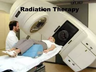
PART III: Particle therapy of cancer. MID 42330. 1. Provide a lethal dose to the tumor. and Spare perfectly the surrounding healthy tissue. The HOLY GRAIL of Radiation Therapy. Ideal Situation. In Practice. Deposit the radiation dose more precisely in the target volume

E N D
PART III: Particletherapy of cancer MID 42330 1
Provide a lethal dose to the tumor and Spare perfectly the surrounding healthy tissue The HOLY GRAIL of Radiation Therapy Ideal Situation In Practice Deposit the radiation dose more precisely in the target volume with less dose in the surrounding healthy tissues.
Extra Dose Dose photons proton Bragg Peak Depth in Tissue Tumor extension Photon-Proton dose distribution comparison The spread out Bragg peak (SOBP) proton SOBP • Position of the Bragg peakdepends on beamenergy • Incommingenergy modulation SOBP • No dose downsteam of tumor • Lower dose upstream of tumor 4
Photon IMRT Photon Proton Particle Therapy: Comparing PT & Conventional RT Main advantage of the Bragg peak => more preciseconformalmapping of the tumor volume Scattering technique IMRT = Intensity Modulated Radio Therapy Conventional Radiotherapy 5
Medulloblastoma Treatment X-Rays vs Protons PROTONS 100 60 10 Low or No Dose Released Here X-RAYS High Dose Released Here 6
Radiobiological effects of ionizing radiation Complex mechanism depending on several factors • Type and state of irradiated cells (O2, SS or DS DNA) • Type and energy of incident radiation (LET) • Quantity of radiation per mass unit (dose) The objective is to break the DNA in the tumorcellsuchthatitcan not repairitself 7
Possible effects of on the cells 1 2 1 => apply fractions 2 or 3 => wanted 4 => not wanted => mayinducesecondarytumors in healthy tissues => avoid by applyingmultiple fractions 4 3 Tumorcells have lessrepaircapability => apply fractions sothathealthycellscanrepair
Relative Biological effect of Protons vs Carbon • LET LinearEnergy Transfer • Fast protons and photons low LET radiation • Carbon, neutrons and slow protons high LET radiation • Low LET • Relativelyuniform distribution of dose • DNA isbroken by oxidizingradicalscreated in water/tissue (indirectly) • High LET • High density of ionizationeventsalongparticletrack • DNA isbroken by direct ionizationevents • OER OxygenEnhancement Ratio • Low LET radiation more effective in a oxygen-richenvironment • Tumors are lessoxygenated (lessvascularized) • Healthy tissue ishigheroxygenated (wellvascularized) • High LET Relative Biologicaleffectiveness (RBE) ishigher
LET: LinearEnergy Transfer Photons, Fast Protons Low LET Neutrons, Carbon ions High LET Dose more defused, lessconcentrated Dose concentrated on track
Two types of radiation damage Indirect: Low LET particles (g,X,fast n, or b) induceradiolysis in the cell. The chemicallyveryreactive free radicals destroy the DNA strand. The presence of O2 increases the production of free radicals, therefore the radio-sensitivity of the cells to low LET radiation Indirect effects are dose and state of cell dependant. Direct:High LET particles (C,α, slow n, p or β) have frequent interaction over a short distance; they break directly DNA strands due to massive but local destruction. Whatever the state of the cell, enzymes generally cannot repair the DNA.
OER A famous experiment of Dr. Gray dose • Cellsurvival rate as function of absorbed dose • Oxygenpoorcondition (tumor) • Normal oxygen condition • OER = 2.9 survival 2.9
OER decreases as the LET increases High -LET OER for high LET = 1 - 1.7 Oxygen Enhancement Ratio Low O2-level in tumors =>Carbonbeamsevenbetterthan proton beams dose Low -LET survival OER for low LET = 3
Ability to reach the tumor Range in patient: up to 32 g/cm² Range modulation:up to full range, with steps of 0.5 g/cm² Field size: up to 30 x 40 cm Ability to reach the from any selected direction Isocentric Gantry Precise, robotic patient positioner Ability to reach the tumor accurately Penumbra: maximum 2 mm at skin Distal dose falloff: maximum 1 mm above physical limit Patient positioner accuracy and reproducibility: 0.5 mm for small displacements Gantry accuracy and reproducibility: 1 mm radius circle of confusion Patient alignment methods: lasers, light fields, X-rays Ability to verify and control the dose deposition using IC’s Main Specifications of a Proton Therapy System
Accelerator parameters driving the technology choice • Energy: defines the range in the patient (230 MeV enough) • Energy definition: defines the range accuracy and the distal falloff • Beam current: defines the dose rate (1011 p/sec enough (10 nA)) • Beam current stability and noise: defines ability to use wobbling and scanning • Accurate and fast beam current control: needed for conformal therapy
Cyclotrons for Proton & Carbon therapy? • In 1991, when IBA entered in PT, the consensus was that the best accelerator for PT was a synchrotron • IBA introduced a very effective cyclotron design, and today the majority of PT centers use the cyclotron technology (Not only IBA but Accel/Varian, Still Rivers Systems/Mevion) • Over these 15 years, users came to appreciate the advantages of cyclotrons: • Simplicity & reliability • Intense, continuous (non pulsed) beam current • Lowest cost and size • But, most importantly, the ability to modulate rapidly and accurately the proton beam current 16
Real oscilloscope measured signals sub-msec response time Modulation signal Beamcurrent 17
Why is fast current modulation important? • A big issue with scanned beam is the motion of the target during irradiation • If you cannot control accurately and rapidly the current ,or if the beam is slowly pulsed , your only choice is step-and-shoot (spot scanning) • Assuming a 10 mm (FWHM) beam spot size, a 50% overlap and a 20 Hz pulse rate, the maximum scanning speed will be 0.2 m/sec • With this speed, for a large size tumor, repainting many time each layer is not really an option • In contrast, with a cyclotron you can scan at 20 m/sec and rescan many times each layer 18
Change of energy? • Cyclotrons are simpler at fixed energy • Energy change by graphite degrader at waist after cyclotron exit, followed by divergence slits and energy analyzer • This very effectively decouples the accelerator from the patient • Fragmentation products are effectively eliminated in slits and ESS • Yes, neutrons are produced, but ESS is well shielded and the average beam currents are very low > little activation • How fast? 5 mm step in energy in 100 msec. Respiration cycle is 2…4 seconds=> 100 msec is fine 19
Small synchrotron Advantages Naturally variable energy Disadvantages Current limited if low energy injection Beam current stability & low noise is difficult on small synchrotrons Fast and accurate beam current control difficult to achieve More complex with negative impact on availability Compact cyclotron Advantages No physical current limitation Beam current stability & noise specifications currently achieved on small cyclotrons Fast and accurate beam current control over 1000/1 range easy to achieve Low complexity, resulting in highest availability Disadvantages Variable energy requires external Energy Selection System Accelerators for proton therapy: two alternatives
A Proton Therapy Facility is like a small Hospital • A proton therapy system is much more than only an accelerator • It is a complex, multi-room system, filling a hospital building. • The total investment is around 100 M€, of which 45 M€ for the equipment • Many people (doctors, therapists, physicists, nurses) work daily in a PT facility • A PT facility can treat 1500 patients/year and generate revenues in the order of 30 M€/year!
Main Sub-systems of a cyclotron based PT facility • 230 MeV isochronous cyclotron • Energy Selection System (ESS) • Beam Transport and Switching System • Isocentric Gantries (typically 3) and one Fixed Beam Line • Nozzles for matching the beam wrtthe required treatment (scattering, wobbling or scanning, diagnostics) • Robotic Patient Positioners • Software Control and Safety System
Typical Proton Therapy Facility Layout control room Fixed beam room accelerator gantry rooms Beam Transport System Experimental area ESS shielding
Proton therapy center €30-55 millions for equipment €45-100 millions for the center 80-100 m 35 m 24
The 230 Mev Cyclotron at MGH in Boston Protons only Fixedenergy 200 tons = 4.7 m
C230 inside view: spiral sectors with elliptical gap Harmoniccoils ESD Spiral sectors => better vertical focusing Elliptical gap => high B-field and easier extraction
IBA PT subsystems : the Cyclone 235 Anotherviewfrom the inside… 28
Extraction from the C230 cyclotron deflector Gradient corrector SmCo doublet
Electrostatic Deflector septum HV electrode
The Energy Selection System • Carbon wedge is used for coarse energy definition • Emittance slits are used to define the emittance of the transmitted beam • Analyzing magnet system defines accurately the range at nozzle entrance • Laminated magnets and quads allow 10% energy change in 2 seconds
More Expertise IBA PT subsystems : the beam transport lines. The energyselection system. WPE, Essen, 2010. 35
More Expertise IBA PT subsystems : the treatment rooms. The isocentricgantry. About 10 m high 37
The purpose of the nozzle • Modulate the proton energy (range in patient) • to spread the proton beam to obtain a uniform dose distribution in a large volume • Double scattering for small to moderate fields • Wobbling for the largest and deepest fields • Pencil Beam Scanning for the most precise conformal mapping • to measure accurately the dose delivered to the patient • Provide alignment of the patient with the proton field
IMPT: Pencil Beam Scanning principle Tumor divided in iso-energy slices Scanning magnets in x and y Y Slices already treated Z X proton beam 2-D dose distribution on the actually scanned slice Bragg-peak
PT center under operation Proton Therapy is growing rapidly ! Courtesy Janet Sisterson & PTCOG 42
Orsay, France More than 20 IBA PT customers in the world Skandion, Sweden MGH, Boston Beijing, China Carl Gustav Carus, Dresden, Germany ProCure2, Chicago NCRH, Cracow, Poland ProCure4, Seattle Procure 3, Somerset, NJ Dimitrovgrad, Russia NCC, Kashiwa, Japan MPRI, Indiana KNCC, Ilsan, Korea ProCure 1 Oklahoma City U.Penn, Philadelphia PTC Czech, Prague ProVision Knoxville, Tennessee Trento, Italy Wanjie, China Hampton Univ., Virginia WPE, Essen, Germany UFPTI, Jacksonville, FL 43
The UPHS Particle Therapy Centre, Philadelphia • The largest Particle Therapy centre to date! • 4 Gantry Rooms • 1 Fixed Beam Room • 1 Experimental Room • Beam since July 2008 45