Download
1 / 59
590 likes | 750 Views
Patient 1. CC: 5YOWFpresents with fever, marked weakness, pallor, bone pain, and bleeding from her nose HPI: progressively increasing fatigability and infections over the past few months

E N D
Patient 1 • CC: 5YOWFpresents with fever, marked weakness, pallor, bone pain, and bleeding from her nose • HPI: progressively increasing fatigability and infections over the past few months • PE: marked pallor; epistaxis; ecchymotic patches over skin; sternal tenderness; slight hepatosplenomegaly with nontender lymphadenopathy; no signs of meningitis; normal funduscopic exam.
Gross Pathology • Neoplastic infiltration of lymph nodes, spleen, liver and bone marrow. • Loss of normal bone marrow architecture.
Micro Pathology • Myelophthisic bone marrow (distorted architecture secondary to a space-occupying lesion) with lymphoblastic infiltration • Lymphoblasts with inconspicuous nucleoli, condensed chromatin, scant cytoplam
Labs • Normocytic, normochromic anemia • Absolute lymphocytosis with excess blasts (>30%) • Negative monospot test for Epstein-Barr virus. • Positive terminal deoxytransferase (TdT)(marker for immature T and B lymphocytes) • Positive CD10 marker (CALLA-Common Acute Lymphoblastic Leukemia Antigen
Diagnosis and Discussion • Diagnosis: Acute Lymphocytic Leukemia (ALL) • Discussion: ALL is the most common pediatric neoplasm; it accounts for 80% of all childhood leukemias. It carries a good prognosis.
Patient 2 • CC: 25YOF presents with high grade fever, menorrhagia, and marked weakness • HPI: recurrent infections over the last few weeks • PE: Marked pallor; multiple purpuric patches over skin; hepatosplenomegaly; gingival hyperplasia; sternal tenderness; normal funduscopic and neurologic exam
Gross Pathology • Bone erosion due to marrow expansion • Chloroma formation, mainly in skull • Splenomegaly
Micro Pathology • Auer Rods (basophilic cytoplasmic bodies) in myelocytes • Peroxidase positive stains on bone marrow and gingival biopsy • Myeloblasts with myelomonocytic differentiation replace normal marrow
Labs • Normocytic, normochromic anemia. • Thrombocytopenia • Neutropenia. • Prolonged PT/PTT • Leukocytosis composed mainly of myeloblasts and promyelocytes
Diagnosis and Discussion • Diagnosis: Acute Myelogenous Leukemia (AML) • Discussion: Not as common as ALL. Increased risk associated with ionizing radiation, benzene exposure, Down’s syndrome, and cytotoxic chemotherapeutic agents
AML/ALL Common Symptoms • Fever, pallor, weakness, bone pain, epistaxis, recurrent infections • Thrombocytopenia; neutopenia • Minor hepatosplenomegaly
Gingival lesions Auer Rods Nontender lymphadenopathy Scant cytoplasm CD10 (CALLA) positive TdT positive AML vs. ALL
Patient 3 • 11Month Old M of Indian descent • CC: Presents with marked pallor, failure to thrive, and delayed developmental motor milestones. • PE: Marked pallor, mild icterus; frontal bossing and maxillary hypertrophy “chipmunk face;” splenomegaly
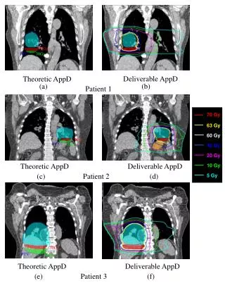
Imaging • XR, Skull (lateral): maxillary overgrowth and widening of diploic spaces with “hair on end” appearance of frontal bone, caused by vertical trabeculae
Gross Pathology • Expansion of hematopoietic bone marrow • Thinning of cortical bone
Micro Pathology • Red marrow increased • Yellow marrow decreased • Marked erythoid hyperplasia in marrow
Labs • Microcytic hypochromic anemia • Decreased reticulocytosis • Mildly increased unconjugated bilirubin • Anisopoikilocytosis • HbA-absent • HbF-95%
Diagnosis and Discussion • Diagnosis: Beta Thalassemia • Discussion: Beta-Thalassemia results from decreased synthesis of beta-globulin chains due to errors in transcription, splicing or translation of mRNA. Alpha-Thalassemia results from decreased synthesis of alpha-globulin chain due to deletion of one or more of the four alpa genes normally present.
Treatment • Blood transfusion, folic acid supplementation, iron chelation therapy, bone marrow transplantation
Patient 4 • 24YOWM • CC: Rapid enlargement of his abdomen, producing a dragging sensation, along with a painless lump in his neck for the past two months • HPI: Intermittent fever, drenching night sweats, pruritus, and significant weight loss • PE: Pallor; unilateral nontender, rubbery, enlarged cervical lymph nodes; splenomegaly; no enlargement of tonsils
Labs • Neutrophilic leukocytosis with lymphopenia • Normocytic anemia • Elevated ESR • Elevated serum Cu and ferritin • Negative Mantoux test
Imaging • CXR: bilateral hilar lymphadenopathy
Gross Pathology • Involved lymph nodes are rubbery • Have “cut-potato” appearance of cut surface
Micro Pathology • Lymph node biopsy shows large histiocyte cells with multilobed nuclei and eosinophilic nucleolus resembling owl’s eyes (Reed-Sternberg Cells) • No bone marrow involvement.
Diagnosis and Discussion • Diagnosis: Hodgkin’s Lymphoma • Discussion: Four patterns seen on biopsy. Common symptoms are the fever, night sweats, and weight loss. The disease spreads to contiguous lymph nodes before moving into the blood.
Patient 5 • 40YOWM • CC: Life insurance physical exam • HPI: No complaints except occasional fatigue and increasing abdominal girth. • PE: Pallor of skin and mucus membranes; markedly enlarged spleen; pain on palpation of sternum; no lymphadenopathy
Gross Pathology • Skull chloromas • Enlarged congested spleen with areas of thrombosis and microinfarcts • Hepatomegaly
Labs • Markedly elevated WBC count (130,000) • Immature granulocytes mixed with normal appearing ones • Basophilia, eosinophilia, early thrombocytosis, late thrombocytopenia • Low leukocyte alkaline phosphatase • Elevated serum vitamin B12 • Chromosomal translocation t(9;22)/bcr-abl gene (Philadelphia Chromosome)
Micro Pathology • Hepatic sinusoidal leukemic infiltrates • Congestive splenomegaly with myeloid metaplasia • Philadelphia chromosome in all myeloid progeny
Diagnosis and Discussion • Diagnosis: Chronic Myelogenous Leukemia (CML) • Discussion: Death usually results from accelerated transformation into acute leukemia (blast crisis) within 2-5 years.
Patient 6 • 65YOWM • CC: Routine checkup • HPI: On directed history, he admits to a weight loss of about 12lbs over the past 4 mos, together with episodes of epistaxis and extreme fatigue • PE: Generalized nontender lymphadenopathy; pallor; enlargement of spleen and liver
Gross Pathology • Lymph node enlargement • Hepatosplenomegaly with tumor nodule formation
Micro Pathology • Bone marrow biopsy reveals extensive infiltration, normal-looking lymphocytes and a few lymphoblasts • Blood smear shows many lymphocytes with small, dark, round nucleus and scant cytoplasm
Labs • Markedly elevated WBC (124,000) • 90% Lymphocytes • No lymphoblasts • Mild thrombocytopenia • Cooms-positive hemolytic anemia • Smudge cells • B-cells express CD5 (normally a T-cell marker)
Diagnosis and Discussion • Diagnosis: Chronic Lymphocytic Leukemia (CLL) • Discussion: CLL is a malignant neoplastic disease of B Lymphocytes that express surface marker CD5. Characterized by slow progression of anemia, hemolytic anemia, recurrent infections, lymph node enlargement, and bleeding episodes.
Patient 7 • 10YOBM • CC: Chronic nonhealing ulcer on lower leg • HPI: Recurrent episodes of abdominal and chest pain along with diminution of vision. • PE: Fever; pallor; mild icterus; funduscopy shows hypoxic spots with neovascularization; nonhealing chronic ulcer on left lower leg
Imaging • CT/US of Abdomen: small, calcified spleen
Labs • Decreased HCT • Megaloblastic anemia • Serum bilirubin moderately elevated • Howell-Jolly bodies and Cabot rings
Diagnosis • Sickle Cell Anemia
Patient Next • 10 Month old Female • CC: Mother claims the child is “a retard.” She cannot see properly, and falls repeatedly. • PE: No lacerations of fractures noted; normal physical development for size and weight; bruises in different stages of healing; bilateral retinal hemorrhages
Imaging and Labs • Imaging: XR: No new or old fractures • Labs: Coagulation profile is normal.