Download
1 / 67
680 likes | 685 Views
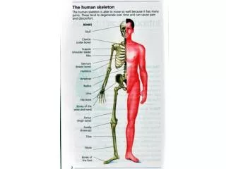
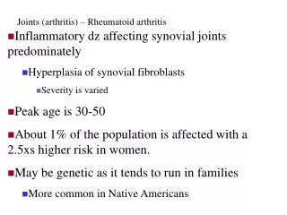
Acute Arthritis. Dr. Müge Bıçakçıgil. Acute Arthritis. The sudden onset of inflammation of the joint, causing severe pain, swelling, and redness. Structural changes in the joint itself may result from persistence of this condition. Signs of Inflammation. Swelling Warmth Erythema

E N D
Acute Arthritis Dr. Müge Bıçakçıgil
Acute Arthritis The sudden onset of inflammation of the joint, causing severe pain, swelling, and redness. Structural changes in the joint itself may result from persistence of this condition.
Signs of Inflammation Swelling Warmth Erythema Tenderness Loss of function
Key Points Distinguish arthritis from soft tissue non articular syndromes If the problem is articular distinguish single joint from multiple joint involvement Inflammatory or non-inflammatory disease Always consider septic arthritis!
INFLAMMATION •Acute/chronic •Monoarthritis •Oligoarthritis •Polyarthitis
Is it acute or chronic? < 6 weeks Acute > 6 weeks chronic Minutes to hours : hemarthrosis Hours to days: septic, reactive, crystals Days to weeks: autoimmune ( RA), CTD, viral Weeks to months: degenerative, other
Acute Monoarthritis Inflammation (swelling, tenderness, warmth) in one joint Occasionally polyarticular diseases can present with monoarticular onset: (RA, JRA,Reactive and enteropathic arthritis, Sarcoid arthritis, Viral arthritis, Psoriatic arthritis)
Acute Monoarthritis - Etiology THE MOST CRITICAL DIAGNOSIS TO CONSIDER: INFECTION ! Septic Crystal deposition (gout, pseudogout) Traumatic (fracture, internal derangement) Other (hemarthrosis, osteonecrosis, presentation of polyarticular disorders)
Joint aspiration must be done! Needed for immediate diagnosis. Bloody joint aspirate- plain X-ray . Analysis of synovial fluid provide discrimination of infection and crystal artropathy.
septic arthritis common organisms Staphylococci or Streptococcus young adults, significant incidence gonococcal arthritis Elderly & immunocompromised gram (-) organisms Anaerobes more common with penetrating trauma
Who gets septic arthritis? pre-existing joint disease prosthetic joints low SE status, IV drug abuse, alcoholism diabetes, steroids, immunosuppression previous intra-articular steroid injection
Symptoms & signs of septic arthritis • Typically hot, swollen, red tender joint with reduced range of movement, difficulty weight bearing • Systemic upset • Night and rest pain • Large joints more commonly affected than small
Symptoms & signs of septic arthritis Delayed or inadequate treatment leads to irreversible joint damage 10-15% of cases, > one joint - so polyarticular presentation does not exclude septic arthritis presence of fever not reliable indicator- if clinical suspicion high - treat
Investigations • Synovial fluid aspiration • volume/viscosity/cellularity/appearance • gram stain/culture • Absence of organism does not exclude septic arthritis • polarised light microscopy (crystals) • suspected prosthetic joint sepsis should ALWAYS be referred to orthopaedics
Septic Joint - Gonococcal Often preceded by disseminated gonococcemia Sexually active individual, fever, chills, skin lesions, migratory arthralgias and tenosynovitis persistent monoarthritis Genitourinary disease often asymptomatic
Tests to Perform on Synovial Fluid Gram stain and cultures . Total leukocyte count/differential: inflammatory vs. non-inflammatory. Polarized microscopy to look for crystals. Not necessary routinely: Chemistry (glucose, total protein, LDH) unlikely to yield helpful information beyond the previous tests.
Investigations Always blood cultures significant proportion blood cultures + ve in absence of + synovial fluid cultures FBC ESR & CRP BUT absence of raised WBC, ESR or CRP not exclude diagnosis of sepsis - if clinical suspicion high always treat
Other tests? If skin pustule is present, suggestive of gonococcal infection, then skin swab should be taken If history suggests possibility of genitourinary or respiratory tract infection then culture sputum (and CXR) & urine & take anogenital & throat swabs where appropriate
Urate crystals Gram positive coccus
Gout Caused by monosodium urate crystals Most common type of inflammatory monoarthritis Typically: first MTP joint, ankle, knee Pain very severe May be with fever and mimic infection The cutaneous erythema may extend beyond the joint and resemble bacterial cellulitis
Stages -Acute intermittent gout Characteristic gout attack: rapid development of warmth, swelling, erythema and pain in the affected joint. The initial attack is monoarticular and in 50% of cases involves the 1st metatarsal joint, which will finally be affected in 90% of patients.
Laboratory Features and Diagnosis Uric acid level in serum is of limited value in establishing the diagnosis: The majority of hyperuricemic subjects will not develop gout. Normal level of uric acid during gouty attack is frequent.
Diagnosis Definitive diagnosis is possible only by aspiration and inspection of the synovial fluid or tophaceous material. Crystals are needle or rod-shaped. On compensated polarized microscopy, they appear as a bright, birefringent crystals (usually intracellular) that are yellow when parallel to the axis of slow vibration.
Urate Crystals • Needle-shaped
Risk Factors Primary gout: Obesity, hyperlipidemia, diabetes mellitus, hypertension, and atherosclerosis. Secondary gout: alcoholism, drug therapy (diuretics), myeloproliferative disorders, chronic renal failure.
Treatment The management of gout involves treating acute arthritic inflammation and urolithiasis lowering urate levels with the goal of preventing recurrent disease and progression.
Treatment of Acute Gouty Arthritis NSAIDs are considered first-line therapy. Selective Cox-2 inhibitors are an alternative in patients with GI contraindications. Corticosteroids or subcutaneous injections of corticotropin are additional alternatives. Because colchicine adverse effects can be serious, IV colchicine should not be used.
Long-Term or Prophylactic Therapy NSAIDs and colchicine are frequently used as prophylaxis against recurrent acute gout, since such episodes are common during the initiation of uric acid–lowering treatment. Allopurinol and Probenecid - apotent uricosuric agents equally acceptable as first-line drug.
CPPD Crystals Deposition Disease Can cause monoarthritis clinically indistinguishable from gout – Pseudogout. Pseudogout is most common in the knee (50%) and wrist. Reported in any joint (Including MTP). CPPD disease may be asymptomatic (deposition of CPP in cartilage).
Ca pyrophosphate (pseudogout) Rod or rhomboid-shaped Weakly positive birefringent
What are other differentials for acute monoarticular pain?
Monoarthritis - differential diagnosis Psoriatic arthritis • Onycholysis • Subungual hyperkeratosis • Pitting • Extensor surfaces, scalp, umbilicus
Monoarthritis - differential diagnosis Reactive arthritis • Prodromal GI /GU Infection eg campylobacter, salmonella, shigella, Yersinia,chlamydia • Pustular psoriasis and circinate balanitis
Monoarthritis - differential diagnosis Trauma and haemarthroses (warfarin, bleeding disorders) Palindromic rheumatism – 24-48 hours inflammatory monoarthritis, can evolve into polyarthritis eg RA
Other Tests Indicated for Acute Arthritis 1. Almost always indicated: Radiograph, bilateral CBC 2. Indicated in certain patients: Cultures PT/PTT ESR 3. Rarely indicated: Serologic: ANA, RF Serum Uric acid level
Acute Inflammatory Oligoarthritis • A patient with 2-4 joints is said to have pauci- or oligoarticular arthritis
Differential diagnosis of acute inflammatory oligoarthritis • Infection • Disseminated gonococcal infection • Nongonococcal septic arthritis • Bacterial endocarditis • Viral • Postinfection • Reactive arthritis • Rheumatic fever (post strep) • Spondyloarthropathy • Ankylosing spondylitis • Psoriatic arthrit • Inflammatory bowel disease • Oligoarticular presentation of RA, SLE, still disease • Gout and pseudogout
Acute Inflammatory OligoarthritisReactive arthritis ( ReA)GIT : Campylobacter, Yersinia, Salmonella, ShigellaGenitourinary: Chlamydia, GonococcusThroat: β hemolytic streptococcus
Acute Inflammatory OligoarthritisPsoriasis associated arthritis ( PsA)Typical joints:DIPs,big and smalltogether
Polyarthritis Inflammation of 5 or more joints Diagnosis cannot always be made with certainty in <6 weeks Bacterial infection less likely but viruses common cause of acute polyarthritis
Acute Polyarthritis Infection Gonococcal Meningococcal Rheumatic fever Bacterial endocarditis Viral (rubella, parvovirus, Hep. B) Inflammatory RA JRA SLE Reactive arthritis Psoriatic arthritis Polyarticular gout Sarcoid arthritis