Download
1 / 17
180 likes | 317 Views
Autonomic Nervous System. OBJECTIVES. At the end of the lecture, students should be able to: Define the autonomic nervous system. Describe the structure of autonomic nervous system Trace the preganglionic & postganglionic neurons in both sympathetic & parasympathetic nervous system.

E N D
OBJECTIVES At the end of the lecture, students should be able to: • Define the autonomic nervous system. • Describe the structure of autonomic nervous system • Trace the preganglionic & postganglionic neurons in both sympathetic & parasympathetic nervous system. • Enumerate in brief the main effects of sympathetic & parasympathetic system
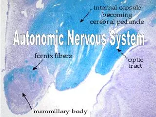
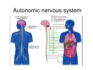
Autonomic Nervous System • Concerned with the innervation and control of involuntary structures: visceral organs, smooth & cardiac muscles and glands • Along with the endocrine system, its primary function is homeostasis of the internal environment • Located both in the central and peripheral nervous systems. • Regulated (controlled)by hypothalamus.
Unlike the somatic nervous system, the efferent pathway of the autonomic nervous system is made up of two neurons called as preganglionic and postganglionic neurons The cell bodies of the preganglionic neurons are located in the brain and spinal cord. Their axons synapse with the postganglionic neurons whose cell bodies are located in the autonomic ganglia Autonomic Nervous System
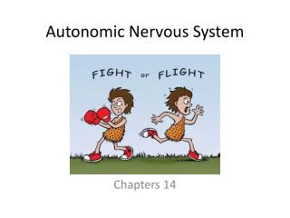
Based on the anatomical, physiological and pharmacological characteristics, the autonomic nervous system is divided into: Sympathetic: Activated during exercise, excitement, and emergencies. “fight, flight, or fright” Parasympathetic: Concerned with conserving energy. “rest and digest” Both divisions operate in conjunction with one another (have antagonistic control over the viscera) to maintain a stable internal environment
Para sympathetic Sympathetic Preganglionic neuron Short Preganglionic fiber Long Ganglion Postganglionic neuron Long Short Postganglionic fiber Target organ Target organ
Sympathetic Division • Preganglionic neurons: located in the lateral gray horn of T1-L2 segments of spinal cord (Thoracolumbar outflow)
Sympathetic Ganglia • Located nearer the central nervous system as: • Prevertebral: celiac & mesenteric • Paravertebralforming sympathetic chain
Paravertebral Ganglia • They are interconnected to form 2 sympathetic chains, one on each side of vertebral column. • Number of ganglia: • Three in cervical part of chain • Eleven to twelve in thoracic part • Four in lumbar & sacral parts each. • The chains end into a common ‘ganglion impar’ in front of coccyx
Preganglionic fibers Run in the ventral roots of the spinal nerve Travel through the spinal nerve, and then join the sympathetic chain via the white ramicommunicans. (WRC)
Within the sympathetic chain, these fibers may: Ascend, descend or remain at the same level to synapse with neurons (postganglionic) of paravertebral ganglia located in sympathetic chain. Leave the sympathetic chain (without synapse) to reach coeliac & mesenteric ganglia(around branches of abdominal aorta) to synapse with their neurons (postganglionic).
Postganglionic fibers From the sympathetic chain ganglia enter again into the spinal nerve through grey ramicommunicantes (GRC)to supply structures in head & thorax + blood vessels & sweat glands From the cells of coeliac & mesenteric ganglia supply abdominal & pelvic viscera.
Parasympathetic Division Preganglionic neurons Located in: • Nuclei of the 3rd, 7th, 9th & 10th cranial nerves, in the brain stem (Cranial outflow) & • The lateral gray horn of S2-S4segments of spinal cord (Sacral outflow)
Parasympathetic Division • Preganglionic fibers from cranial outflow are carried by 3rd, 7th, 9th & 10th cranial nervesand terminate in ciliary, pterygopalatine, submandibular, otic & peripheral ganglia • Postganglionic fibers innervate organs of the head, neck, thorax, and abdomen
Parasympathetic Division • Preganglionic fibers from sacral outflow are carried by pelvic splanchnic nerves to peripheral ganglia in pelvis where they synapse. • Postganglionic fibers innervate organs of the pelvis and lower abdomen