Download
1 / 53
610 likes | 701 Views
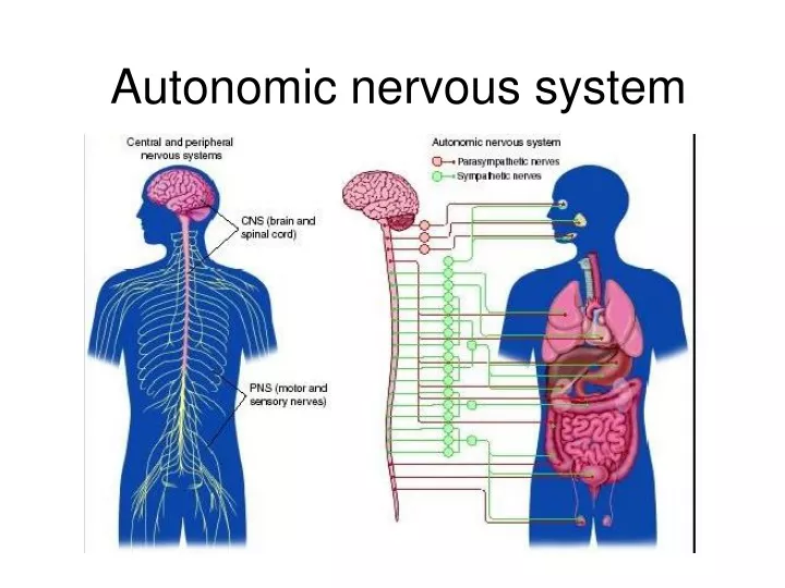
Autonomic nervous system. AUTONOMIC NERVOUS SYSTEM. autonomic nervous system participates in innervation of the visceral part of body, it controls autonomic functions, which takes place independently of our will It is consist of visceromotor nerve fibers

E N D
AUTONOMIC NERVOUS SYSTEM • autonomic nervous system participates in innervation of the visceral part of body, it controls autonomic functions, which takes place independently of our will • It is consist of visceromotor nerve fibers • It makes sensory innervation of visceral organs, vessels, motor innervation of smooth muscle and myocardium and glandular cells • It includes neurons of CNS and PNS • central part – hypotalamus, reticular formation medulla oblongata, spinal cord, cortex • peripheral part – nerve fibers (cranial nerves, spinal nerves)
Types of stimuli Nuclei within CNS visceromotor fibers – through anterior roots of spinal cord autonomic ganglia along the spine – to the organs of abdomen, thorax, pelvis Free nerve endings in the wall of organs pressure, thrust, pain- viscerosensory autonomic ggl.- to posterior roots of spinal cord - ggl. spinale or ggl. VII., IX., X. and into visceromotor nuclei
Autonomic tracts don´t go from CNS directly – they switch over in ganglia outside CNS They are formed at least by two neurons, which switch over in so-called autonomic ganglion Preganglionic neuron : myelinated axon that goes from CNS to autonomic ganglion Postganglionic neuron: unmyelinated axon that goes from autonomic ganglion as a proper autonomic nerve
autonomic (visceromotor) nerve fibers are oftwotypes sympatheticparssympathica parasympatheticparsparasympathica Glands and smoothmuscleofalmosteachvisceral organ are innervated by bothsympathetic and parasympathetic Onesystemisusuallyactivating and theotherinhibiting Exception are smoothmuscleofthe skin and skin glands, which are innervatedonly by sympathetic
Main functions Contraction and relaxation of smooth muscle Function of all exocrine and some endocrine glands Hearth rhytm Some metabolic processes
Division of autonomic nerve system • sympathetic – fight or flight • parasympathetic – rest or digest • enteric system
Pars sympathica: nuclei in CNS and in the spinal cord (C8 – L3) Pars parasympathica: nuclei in CNS (which belong to the cranial nerves), spinal cord (S2 – S4) craniosacralsystem (parasympathetic) thoracolumbarsystem (sympathetic) cranio-sacralsystem (parasympathetic)
Sympathetic and parasympathetic system differ in the arrangement of ganglia: Sympathetic ganglia: • are far from target organs (at spine) – paravertebral ganglia – truncus sympathicus dexter et sinister Parasympathetic ganglia: • closer to organs (ganglion ciliare, pterygopalatinum, oticum, submandibulare + scattered within organ walls) Mediators of sympathetic and parasympathetic system: • preganglionic – the same (from CNS) – acetylcholine • postganglionic - sympathetic – noradrenalin • postganglionic - parasympathetic - acetylcholine
SYMPATHETIC „thoracolumbar system“
Arises from the thoracic and lumbar parts of the spinal cord – from nucl. intermediolateralis C8-L3- so-called thoracolumbar system it leaves the spinal nerve as ramus communicans albus - it ends in sympathetic ganglion next to the spine - preganglionic section - to paravertebral ganglia single paravertebral ganglia form truncus sympathicus from the ganglia arise proper sympathetic nerves, postganglionic section Sympathetic nerves enter through different way the innervated organs
It controls the catabolic functions, activates functions of the visceral organs it accelerates the heart activity and breathing It causes contraction of smooth muscle of vessels within the skin and visceral organs and thereby increases blood pressure It increases level of sugar in blood It expands pupils (mydriasis) It conversely slows digestion It induces a state of wakefulness ans it is used in stress reactions Functions
Truncus sympathicus ganglion trunci sympathici (21-25) = paravertebral ganglia rr. intergaglionares rr. communicantes albus + griseus rr. vasculares – periarterial plexuses rr. viscerales=nn. splanchnici - to prevertebral ganglia Cervical part Thoracic part Abdominal part Pelvic part
Ganglion cervicale superiusGanglion cervicale medium Cervical part Ganglion cervicothoracicum / stellatum - It forms periarterial plexuses around a. carotis ext. et int. – intake of sympathicus to neck and head - nn. cardiaci – innervation of the heart
Ganglia thoracica(thoracic part) • 10 pairsof ganglia • nn. splanchnici – forsmoothmuscleof GIT anditsvessels • rr. communicantesgrisei - to intercostalnerves • Branches to heart, lungs, esophagus • Ganglia lumbalia(lumbar, abdominal part) • 4-5 pairsof ganglia • rr. communicantesgrisei • nn. splanchnicilumbales • rr. vasculares • Ganglia sacralia(pelvic part) • 4 pairsof ganglia • rr. communicantesgrisei – forpelvicorgans • Periarterialplexuses
Prevertebral ganglia and plexuses • They are formed by fibers arising from paravertebral ganglia • On the anterior wall of abdominal aorta • Mixed plexus– nn. splanchnici + n. vagus
pars cranialis- III.,VII.,IX.,X. (cranial parasympathetic) pars sacralis S2-S4 (sacral parasympathetic) - craniosacral system - ganglia are located close to the innervated organs, preganglionic section is therefore long and postganglionic section is short mediator is acetylcholin in whole section- cholinergic system
pars cranialis: most important is parasympathetic part of nervus vagus – it innervates the digestive tract till the border between colon transversum and colon descendens in the abdominal cavity pars sacralis: it innervates the digestive tract from the border between colon transversum and colon descendens till rectum and visceral organs located in the pelvis (urinary bladder, genital organs except gonads) Functions It controls anabolic reactions – preservation of energy, it induces inhibition of organism: - It slows heart activity and breathing • It decreases blood pressure • It narrows pupils (miosis) • It accelerates digestion, sweating and salivation • It is used especially ar rest (slep) and during digestion
Parasympathetic = craniosacral system • Nuclei of cranial nerves: ncl. oculomotorius accessorius to ganglion ciliare (m. sphincter pupilae, m. ciliaris) ncl. salivatorius superior (VII.) to ganglion pterygopalatinum and submandibulare (lacrimal gland, mucosa of nasal cavity, palate, tongue, gl. sublingualis and submandibularis) ncl. salivatorius inferior (IX.) to ganglion oticum (glandula parotis and small salivatory glands of cheek) ncl. dorsalis n. X (together with n. vagus to organs) • ncl. intermediolateralis S2-4 (pars sacralis, pelvica) – to pelvic organs nn. splanchnici pelvici ganglia are located within the skull or organs walls
Ganglion ciliare • Here end preganglionic fibers of n.oculomotorius • parasympathetic (m. sphincter pupillae, m. ciliaris) sympathetic (m. dilatator pupillae) Ganglion pterygopalatinum • Here end preganglionic fibers of n.facialis • Mucosa of posterior part of nasal cavity, upper teeth, mucosa of hard palate, lacrimal gland Ganglion submandibulare • Here end preganglionic fibers of n.facialis • gl. sublingualis, gl. submandibularis, salivary glands of tongue and botoom of oral cavity Ganglion oticum • Here end preganglionic fibers of n.glossopharyngeus • skin, mucosa, teeth and gingiva of lower jaw, gl. parotidea Preganglionic fibers of n. vagus end in prevertebral ganglia of thoracic and abdominal cavity Preganglionic fibers of sacral parasympathetic are switched over in pelvic plexuses
Entericsystem • In the wall of digestive tract • plexus submucosus • plexus myentericus • Separate and independent of connection with sympathetic and parasympathetic • It works also after interruption of connections with ANS • It controls tension and mobility of digestive tract, it regulates secretion of all glands and blood flow • innervation and regulation of function of gall bladder and pancreas
CNS The highest autonomic headquarters= hypothalamus It is controled by limbic system
and viscerosensory
THE SENSORY TRACTS • receptor→CNS • A) specific:specificinformation • B) nonspecific: throughinterneurons, generalinformation, preparationof CNS forincomeofspecificinformation • 1th neuron: pseudounipolar cell of spinal ganglion (ganglion ofcranialnerves) →(cerebellum)→thalamus→cortex • SOMATOSENSORY TRACTS: protopathic sensibility epicritic sensibility proprioception • VISCEROSENSORY TRACTS
Protopathic sensibility: tactileinformation (warmth, cold, pressure, pain, rough skin sensibility) • Limbsand trunk: tractus spino-thalamo-corticalis 1stPseudounipolar neuron of spinal ganglion→2ndnucleus proprius→3rd thalamus →cortex (gyruspostcentralis, area 1, 2, 3) 2) Head area: tractustrigemino-thalamo-corticalis 1st Pseudounipolarneuronsof sensory ganglia of CN (V., VII., IX., X.) → 2ndnucleustractusspinalis (V.) → 3rdthalamus→cortex
Epicritic sensibility: discriminatorysensation (tactileresolutionofshapeofobjectetc.) • Limbsand trunk: tractus spino-bulbo-thalamo-corticalis 1st Pseudounipolar neuron ofspinalganglion→fasciculusgracilis, fasciculuscuneatus → 2nd nucleusgracilis, cuneatusmedialis→ 3rd thalamus →cortex (gyruspostcentralis, area 1, 2, 3) 2) Head area: tractustrigemino-thalamo-corticalis 1st Pseudounipolarneurons od sensory ganglia of CN (V., VII., IX., X.) → 2nd nucleusprincipalis (V.) → 3rd thalamus→cortex
Proprioception: fromthelocomotorsystem to the cerebellum • LL and trunk: 1stPseudounipolar neuron od spinal ganglion→ 2ndnucleusthoracicus→ 3rd cerebellum →4th thalamus → cortex 2) UL: 1st Pseudounipolar neuron of spinal ganglion →fasciculuscuneatus → 2nd nucleuscuneatuslateralis → 3rd cerebellum → 4th thalamus→cortex
3) Head area: tractustrigemino-thalamo-corticalis 1stPseudounipolarneuronsofnucleusmesencephalicus nervi V. → 2nd cerebellum → 3rd thalamus→cortex • VISCEROSENSORY TRACTS 1st Pseudounipolar neuron of spinal ganglion→ 2ndnucleusintermediomedialis → nucleusintermediolateralis → FR →thalamus →cortex
MOTOR TRACTS • Set of all neural tracts, which are are connected into the regulation of movement. To them belong pyramidal and extrapyramidal tracts.
PYRAMIDAL TRACTS (direct) • projection direct motor tracts of voluntary movement • They interconnect motor cortex of hemisphere with motoneurons of anterior spinal horns and with motoneurons of nuclei of cranial nerves • It is only one-neuron way • They start in primary motor cortex, to them belong tractus cortico-spinalis (tract of voluntary movement of trunk and limbs) and tractus cortico-nuclearis (tract of voluntary movement of striated muscles of the head ).