Download
1 / 28
280 likes | 403 Views
M267 Lecture 3, Cell Cycle February 24, 2006. G 0. Growth factor/ Mitogen. Restriction point. G 1. M. G 2. S. c. RB. RB. Cyclin D1. p. Mitogen signal for G 0 cells. Cell cycle progression. CDK 4. E2F. E2F. Late G 1 , Early S. G o , Early G 1.

E N D
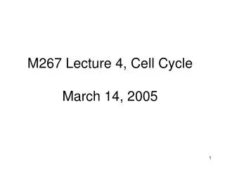
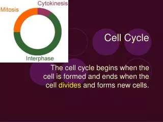
M267 Lecture 3, Cell Cycle February 24, 2006
G0 Growth factor/ Mitogen Restriction point G1 M G2 S
c RB RB Cyclin D1 p Mitogen signal for G0 cells Cell cycle progression CDK 4 E2F E2F Late G1, Early S Go, Early G1 C-myc, cyclin E, B-myb, E2F-1, DNA polymerase alpha, thymidine kinase, thymidylate synthase, histones H2A, H2B,dihydrofolate reductase
Cyclin D1 cdk4 Cyclin B G1 M cdc2 G2 S
H2A H2B H1
NPAT NPAT p p p p NPAT p p H2A H2B H1 Cyclin E Cdk2 + ATP + ATP + ATP
Fold Induction H4 H4-1 Control
CyclinAHA cdk2 w.t. hpm Histone H1 p107 E2F
inactive T14Y15 T14Y15 T14Y15 T14Y15 T14Y15 T14Y15 T14Y15 T14Y15 T14Y15 T14Y15 T14Y15 T14Y15 T14Y15 T14Y15 T14Y15 T14Y15 T14Y15 T14Y15 T14Y15 T14Y15 T14Y15 T14Y15 T14Y15 cdc2 cdc2 cdc2 cdc2 cdc2 cdc2 cdc2 cdc2 cdc2 cdc2 cdc2 cdc2 cdc2 cdc2 cdc2 cdc2 cdc2 cdc2 cdc2 cdc2 cdc2 cdc2 cdc2 Cycin B is degraded G1 G1 G1 G1 G1 G1 T161 T161 T161 T161 T161 T161 T161 T161 T161 T161 T161 T161 T161 T161 T161 T161 T161 T161 T161 T161 T161 T161 T161 S S S (inactive) M M M G2 G2 G2 p p p p p p p p (still inactive) Active CDK!! ATP Cyclin B Cyclin B Cyclin B Cyclin B Cyclin B Cyclin B Cyclin B Cyclin B Cyclin B Cyclin B Cyclin B Cyclin B Cyclin B Cyclin B Cyclin B Cyclin B Cyclin B T14 dephosphorylation Y15 dephosphorylation PPase
wee1 cdc 25 Cdc2 p34 Mik 1 cdc 13 (Cyclin) Interphase mitosis G2 M
p34 p34 p p p p Histone Histone cyclin Wee 1 Cdc 25 cyclin Histone Histone
Cdc 25 Cdc 25 cdc2 cdc2 cdc2 Cyclin Cyclin Cyclin B B B p p p p p PPase Wee 1 Histone mitosis + ATP Histone
p cdc28 p Clb2 Histone Histone
SIC 1 SIC 1 cdc28 cdc28 p p Clb2 Clb2
p SIC 1 cdc28 p Clb2 Histone Histone
p SIC 1 SIC 1 cdc28 cdc28 p p Clb2 Clb2 Histone Histone
Cdc28/Cln2 Cdc28/Clb2 pET pETSIC1 pET pETSIC1 Hist H1 Purified Sic1 inhibits the Clb2-associated kinase, but not the Cln2 kinase in in vitro. HisHASic1 (pETSIC1) was added to yeast extracts from CLB2HA3 orGAL-CLN2HA3. After 30 min. incubaton at 4oC, Clb2 or Cln2, Sic1 and their associated protiens were immunprecipitated with 12CA5 Antibody, and H1 kinase activity was measured. In control lanes (pET), E. coli extracts from cells containing the empty vector were added.
SIC 1 SIC 1 cdc28 cdc28 p p Clb2 Clb2
p SIC1 must be degraded for cells to enter S SIC 1 SIC 1 cdc28 cdc28 p p Clb2 Clb2 Histone Histone (Don’t be fooled by the use of H1 as a convenient substrate)
CKI CDK CKI CDK p p cyclin cyclin Active CDK Inactive CDK
a-factor Far1
Far1 cdc28 Far1 cdc28 p p cln1 cln1 Active CDK Inactive CDK
Cdc28Cln1 Cdc28Cb2 Cdc28Cb5
p p p p p p p p p cdk cdk WEE1 cyclin Phosp. Thr 161 cyclin WEE1 Dephosp. Thr 161 p cdc25 p cdk cdc25 cyclin ACTIVE CDK Degrade cyclin CKI CKI cdk cdk p cyclin
Pick up a copy of the handout as you leave; please read it before Monday’s lecture