Download
1 / 30
300 likes | 397 Views
Bone Injuries - Traumatic. 1. Very High load (low frequency) 2. Unusual type of load – One which the skeletal structure isn’t designed to handle. 3. Combination loads – Bending stress, loads from various directions, etc. Bone Injuries - Fatigue.

E N D
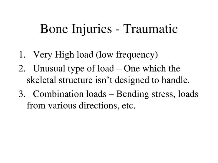
Bone Injuries - Traumatic 1. Very High load (low frequency) 2. Unusual type of load – One which the skeletal structure isn’t designed to handle. 3. Combination loads – Bending stress, loads from various directions, etc.
Bone Injuries - Fatigue • 1. Material fatigue or overuse or "stress fracture" • 2. Very High frequency (moderate to high loads) • 3. Nutritional and hormonal factors increase risk • ex. low Ca2+ intake, low estrogen levels
Fractures • avulsion (tensile) – Often accompanies tendon and ligament injuries. • spiral (torsion) • impacted (compression or crush) • greenstick fracture – Usually seen in children whose bones are not fully mineralized. • comminuted – The bone is broken into several pieces • fatigue or stress fracture
Avulsion fracture of the origin of the sartorius muscle on the iliac spine
Comminuted Fractures Radius Tibia
Stages of Rehabilitation after Bone Fracture • set fracture, immobilize – Allow bone to reach a stable state • reconditioning – Return the bone to its full strength • Therapeutics after fracture • goal: quick restoration of normal function
Fracture Healing • Unlike other tissues, bone heals by regeneration or replacement with the same type of tissue • No scar formation • Original physical integrity and biomechanical properties may be regained
Fracture Healing Sequence • Inflammatory phase (1-5 days) • Swelling, increased blood flow signal • Hematoma (blood clot) forms • prostaglandins, histamine, free radicals (nitric oxide, hydrogen peroxide, etc.) released • WBC's, platelets released • growth factors released • Insulin-like growth factor (IGF-1) • Transforming growth factor (TGF)
Fracture Healing Sequence • Inflammatory phase (continued) (1-5 days) • The clotted hematoma is converted to granulation tissue • New capillary network forms (angiogenesis) • Fibroblasts (fibrous connective tissue), chondroblasts (cartilaginous tissue) and osteoblasts (bone cells) are released
Fracture Healing Sequence • B) Reparative phase • Granulation tissue matures into fibrocartilage mass which holds bone fragments together • Bone callus forms (6 – 12 weeks) • Soft callus – fibrous tissue cartilaginous tissue • Hard callus – cartilaginous tissue bonelike tissue • Clinical union completed (12 – 16 weeks)
Fracture Healing Sequence • C) Remodeling phase (2 months to several years) • callus shrinks • Bone regains original material properties
NOTE: The illustrations and information on the following six slides are courtesy of Scott J. Hollister, PhD – Dept of Biomedical Engineering – University of Michigan
CHANGES IN BONE TISSUE DURING HEALING Remodeling Callus Formation Hardness Bone resorbed by osteoclasts and replaced by osteoblasts Time
Mechanical Effects on Fracture Healing • Rigid Fixation • Broken bone segments are rigidly held together • May bypass the formation of a callus • Called primary healing • Non-Rigid Fixation (cast) • Broken bone is stabilized but fragments aren’t pressed together • Callus forms • Called secondary healing
Mechanical Fixation • Metal devices are attached to the bone. • Designed to hold or force the bone fragments together
Internal Plate Fixators Better and faster healing generally seen with plate fixators.
Intermedullary Rods Generally used with fractures of the femur.
Electromagnetic Fields • Low level electromagnetic fields may promote release of growth factors • Some theorize that excessive application may cause abnormal growth (tumors)
Ultrasound • Low intensity ultrasound may promote fracture healing • Promotes increased blood flow • Seen to increase incorporation of Ca++ in cultures of cartilage and bone cells
Immobilization • reduces mechanical stress around area of bone which has suffered fracture • plaster and fiberglass casts (non-removable) weaken bone overall • experience in wartime led to discovery that active recovery shortens healing time
Osteoporosis • Loss of bone mass due to demineralization (calcium loss) • Weakening of bone increases susceptibility to fracture.
Normal Bone Bone With Osteoporosis
Common Sites of Fractures With Osteoporosis • Neck of Femur • Vertebrae >Dowager's hump – Associated with compression fractures of the vertebral bodies • Distal Radius