Download
1 / 21
230 likes | 452 Views
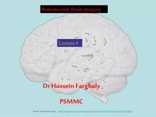
Radionuclide Brain Imaging. Lecture 2. Dr Hussein Farghaly PSMMC. Master Watermark Image: http://williamcalvin.com/BrainForAllSeasons/img/bonoboLH-humanLH-viaTWD.gif. Syllabus Contents. Cerebral Anatomy Cerebral Perfusion Imaging Radiopharmaceuticals Methodology Dosimetry

E N D
Radionuclide Brain Imaging Lecture 2 Dr Hussein Farghaly PSMMC Master Watermark Image: http://williamcalvin.com/BrainForAllSeasons/img/bonoboLH-humanLH-viaTWD.gif
Syllabus Contents Cerebral Anatomy Cerebral Perfusion Imaging Radiopharmaceuticals Methodology Dosimetry Clinical Applications Cisternography Radiopharmaceuticals Methods Pharmacokinetics Dosimetry Clinical Applications
Protects brain from potentially toxic substances from metabolic or dietary substances. Movement across the BBB is controlled by active transport Injury to brain caused by disease, trauma, or toxins causes BBB to lose integrity. Many radiopharmaceuticals depend on specfic transport process BBB Blood Brain Barrier.
BBB imaging useful for: primary or metastatic disease, intracranial inflammation, cerebrovascular disease, complications from head trauma. BBB imaging mainly done today to determine brain death. MRI or CT have taken much of the brain imaging. BBB imaging
Radiopharmaceuticals used to perform BBB imaging do not normally cross the BBB Several Tc99m labeled radiopharmaceuticals can be used to perform BBB imaging. Tc99m (least desirable) accumulates in the choroid plexus, potassium perchlorate is administered, acting as a blocker BBB imaging
Tc99m pentetate-DTPA and Tc99m gluceptate-GH better for planar (static) imaging DTPA/GH do not require pre-treatment of potassium perchlorate. Flow, Blood pool, and static imaging is typically done for BBB imaging. 15-30mCi of Tc99m labeled agent is administered BBB imaging
Symmetric distribution of radiotracer right and left carotid arteries Visualization of superior sagittal sinus Normal delay, symmetric uptake around entire skull BBB imaging
Abnormal distribution Disruption in BBB from lesion, will increase uptake , and localize in area of pathology. Brain death, flow in carotids, complete absence of perfusion in middle and anterior cerebral arteries BBB imaging
Lipid soluble, crosses the bbb, binds to receptors, 92% extraction, 5% brain, 33% lung, liver 44%, dose 3 mCi, give Lugol’s solution, no longer available in the US 123I isopropyl p iodoamphetamine (IMP)
Brain SPECT and PET Radiopharmaceuticals, cont. • Several radiopharmaceuticals are commercially available for brain perfusion SPECT. The most widely used radiopharmaceuticals for rCBF SPECT are 99mTc-labelled compounds, ethyl cysteinedimer (ECD, Neurolite) and hexamethyl propylene amine oxime (HMPAO, Ceretec). • Differences between the two commercially available radiopharmaceuticals, ECD and HMPAO include in vitro stability, uptake mechanism, cerebral distribution and dosimetry. • In normal brain tissue, the kinetic properties of the two agents are very similar. They enter the brain cells due to their lipophilic nature and remain there due to conversion into hydrophilic compounds. • For ECD retention, de-esterification is the crucial reaction leading to hydrophilic conversion, while for HMPAO, instability of the lipophilic form and glutathione interaction have been proposed. • Differences in the retention mechanisms may account for some different behavior of the tracers in specific disorders such as subacute stroke, where ECD distribution seems to reflect metabolic activity more closely, whereas HMPAO is better correlated with cerebral perfusion . As a consequence, both tracers can be used, but they are not interchangeable.
Brain SPECT and PET Radiopharmaceuticals, cont. • For ECD retention, de-esterification is the crucial reaction leading to hydrophilic conversion, while for HMPAO, instability of the lipophilic form and glutathione interaction have been proposed. • Differences in the retention mechanisms may account for some different behavior of the tracers in specific disorders such as subacute stroke, where ECD distribution seems to reflect metabolic activity more closely, whereas HMPAO is better correlated with cerebral perfusion It has to be kept in mind that with the techniques used in clinical practice, ECD and HMPAO SPECT do not provide absolute quantitative flow values but rather estimate relative regional flow differences based on the comparison of count density ratios between various regions (e.g. right/left asymmetries, ratio in relation to reference regions, etc.).
Brain SPECT and PET Radiopharmaceuticals, cont. Comparison between HMPAO and ECD
Brain SPECT and PET Radiopharmaceuticals, cont. Radiation Dosimetry
Brain SPECT and PET Radiopharmaceuticals, cont. • Preparation of the radiopharmaceutical: – Use pertechnetate from generators which have been eluted within the last 24 h. – Use fresh generator eluates not older than 2 h, particularly for HMPAO. – For HMPAO: inject the tracer no sooner than 10 min after radioligand reconstitution. • Quality control Radiochemical purity should be determined on each vial prior to injection using the methods outlined in the package inserts. It should be >90% for ECD and >80% for HMPAO.
Brain SPECT and PET Radiopharmaceuticals, cont. Time interval for injection • Inject the radiopharmaceuticals after quality control check, but not later than 30 min after reconstitution for unstabilized 99mTc -HMPAO, 4 h for stabilized 99mTc –HMPAO, and 6 h for 99mTc -ECD. • Administered activity • – Adults: 555–1110 MBq (15 - 30 mCi) (typically 740 MBq) of either radiopharmaceutical. • - Children : 7.4 -11.1 MBq/kg (0.2 - 0.3 mCi/kg). Minimum dose is 110 MBq (3 mCi)
Brain SPECT and PET Radiopharmaceuticals, cont. • Time from injection to start of data acquisition -Try always to keep the same time delay from injection to the start of data acquisition. – 99mTc-ECD: For best image quality allow a delay of 30–60 min since wash-out from no-specific uptake improves the signal to noise ratio in this period. – 99mTc-HMPAO: For best image quality allow a delay of 30–90 min. – Imaging should be completed within 4 h after injection. Excessive delay should be avoided because of radioactive decay.
Procedure C. Precautions: Continuous supervision of the patients during the whole scanning procedure is necessary. This is especially important for patients with epilepsy and dementing disorders.