Download
1 / 1
10 likes | 138 Views
What’s Your Diagnosis? Abdominal Distention in a Colony of Congenic Mice Eden V. Paster, DVM ; Debra L. Hickman, DVM, MS, DACLAM VA Medical Center, Portland, OR. www.cottontimer.com/2006/08/. Microscopic Findings. Diagnostics

E N D
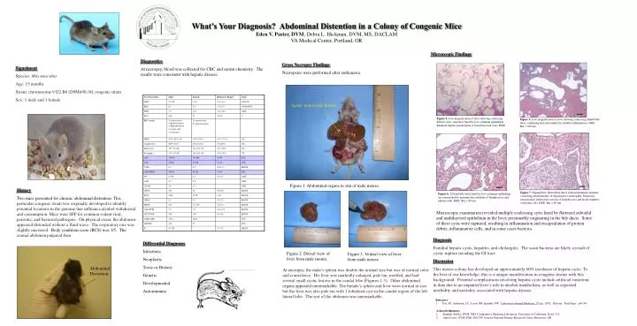
What’s Your Diagnosis? Abdominal Distention in a Colony of Congenic Mice Eden V. Paster, DVM; Debra L. Hickman, DVM, MS, DACLAM VA Medical Center, Portland, OR www.cottontimer.com/2006/08/ Microscopic Findings Diagnostics At necropsy, blood was collected for CBC and serum chemistry. The results were consistent with hepatic disease. Gross Necropsy Findings Necropsies were performed after euthanasia. Signalment Species: Mus musculus Age: 15 months Strain: chromosome 9 D2.B6 [D9Mit90,18] congenic strain Sex: 1 male and 1 female Liver: note cystic lesions Figure 4 Figure 4. Low magnification of liver showing coalescing dilated cystic structures lined by low columnar epithelium. Remnant hepatic parenchyma is found between cysts. H&E. Figure 5. Low magnification of liver showing coalescing dilated bile ducts containing and surrounded by variable inflammation. H&E. Bar = 200 um www.ornl.gov/.../v37_3_04/article08.shtml Figure 1. Abdominal organs in situ of male mouse. History Two mice presented for chronic abdominal distention. This particularcongenic strain was originally developed to identify potentiallocations in the genome that influence alcohol withdrawal and consumption.Mice were SPF for common rodent viral, parasitic, and bacterialpathogens. On physical exam, the abdomen appeared distended without a fluid wave. The respiratory rate was slightly increased. Body condition score (BCS) was 3/5. Thecranial abdomen palpated firm. Figure 7: Hyperplastic dilated bile ducts with intraluminal exudates consisting predominantly of degenerative neutrophils. Periportal mononuclear infiltration consists of lymphocytes and lesser numbers of plasma cells. H&E. Bar = 40 um Figure 6. Dilated bile ducts lined by low columnar epithelium are surrounded by mononuclear infiltrate of lymphocytes and plasma cells.H&E. Bar = 40 um. Microscopic examination revealed multiple coalescing cysts lined by flattened cuboidal and multilayered epithelium in the liver, presumably originating in the bile ducts. Some of these cysts were ruptured, resulting in inflammation and encapsulation of protein debris, inflammatory cells, and in some cases bacteria. Diagnosis Familial hepatic cysts, hepatitis,and cholangitis. The scant bacteria are likely a result of cystic rupture invading the GI tract. Discussion Thismouse colony has developed an approximately 60% incidenceof hepatic cysts. To the best of our knowledge, this is a uniquemanifestation in congenic strains with this background. Potential complications involving hepatic cysts include artificial variations in data due to an impaired liver’s role in alcohol metabolism, as well as expected morbidity and mortality associated with hepatic disease. Differential Diagnoses Infectious Neoplastic Toxic or Dietary Genetic Developmental Autoimmune Figure 2. Dorsal view of liver from male mouse. Figure 3. Ventral view of liver from male mouse Abdominal Distention At necropsy, the male’s spleenwas double the normal size but was of normal color and consistency.His liver was markedly enlarged, pale tan,mottled, and had several small cystic lesions in the caudal lobe (Figures 1-3).Other abdominal organs appeared unremarkable. The female’s spleen and liver were normal in size but the liver was also pale tanwith 1 lobulated cyst in the caudal region of the left lateral lobe.The rest of the abdomen was unremarkable. www.fotosearch.de/ARP112/mouse/ www.nal.usda.gov/.../v8n3/8n3mfg1.htm • Reference • Fox, JG; Anderson, LC; Loew, FM; Quimby, FW. Laboratory Animal Medicine, 2nd ed. 2002. Elsevier. San Diego. p43-44. • Acknowledgements • Stephen Griffey, DVM, PhD; Comparative Medicine Laboratory, University of California, Davis, CA • Anne Lewis, DVM, PhD, DACVP; Oregon National Primate Research Center, Beaverton, OR