Download
1 / 22
230 likes | 331 Views
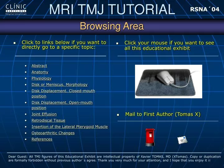
RSNA´04. Browsing Area. Click your mouse if you want to see all this educational exhibit. Click to links below if you want to directly go to a specific topic: Abstract Anatomy Physiology Disk or Meniscus. Morphology Disk Displacement. Closed-mouth position

E N D
RSNA´04 Browsing Area • Click your mouse if you want to see all this educational exhibit • Click to links below if you want to directly go to a specific topic: • Abstract • Anatomy • Physiology • Disk or Meniscus. Morphology • Disk Displacement. Closed-mouth position • Disk Displacement. Open-mouth position • Joint Effusion • Retrodiscal Tissue • Insertion of the Lateral Pterygoid Muscle • Osteoarthritic Changes • References • Mail to First Author (Tomas X) Dear Guest: All TMJ figures of this Educational Exhibit are intellectual property of Xavier TOMAS, MD (XTomas). Copy or duplication are formally forbidden without previous author´s agree. Thank you very much for your attention, and I hope that you enjoy it ¡¡
RSNA´04 Abstract • LEARNING OBJECTIVES • To describe the basic concepts of temporo-mandibular joint MR imaging. • To review the role of MR imaging in the assessment of temporo-mandibular joint dysfunction. Special emphasis is placed on new indirect signs of dysfunction. • To correlate MR imaging features with clinical symptoms. • ABSTRACT Dysfunction of the TMJ is a frequent disease which, in some studies, may affect up to 28 % of the population. In recent years, MRI has been confirmed as the imaging technique of choice in the study of TMJ meniscal displacement in patients. Despite of, a high frequency of disk displacement appears in asymptomatic volunteers. Further studies using the latest techniques allow a better understanding of the sources of joint pain and the discrepancy between imaging findings and patient symptoms. We have systematically analyzed clinical symptoms and MRI signs of TMJ dysfunction which have been previously developed in other studies, such as disk morphology, articular effusion or osseous degenerative changes, as well as other variables not reported, such as the thickness of the insertion zone of external pterygoid muscle and the rupture of retrodiscal ligaments, which may have an important role in the evolution of TMJ dysfunction before osteoarthritic changes lead to a more advanced stage of the dysfunctional spectrum.
RSNA´04 Anatomy 1. Condyle 2. Temporal Bone. Articular eminence 3. Temporal bone. Mandibular fossa 4. Disk. Anterior band (AB) 5. Disk. Intermediate zone (IZ) 6. Disk. Posterior band (PB) 7. Bilaminar zone. Superior retrodiscal layer 8. Bilaminar zone. Inferior retrodiscal layer 9. Bilaminar zone. Vasculo-nervous structures 10. Capsular superior attachment 11. Capsular inferior attachment 12. Superior joint space 13. Inferior joint space 14. Lateral superior pterygoid muscle (SLP) 15. Lateral inferior pterygoid muscle (ILP) 16. Interpterygoid space 17. External auditory canal
RSNA´04 Physiology Initial closed-mouth position (2A) Beginning of the open-mouth position (2B). Digastric muscle forces condylar drop. The condyle rotates in the lower joint space. Afterthat, condylar displacement begins when jaw is opened beyond to 20-25 mm. Retrodiscal ligaments stabilize the disk. Condylar protraction. Maximum open-mouth position (2C). ILP muscle is basic in this step, and SLP can displace the disk, probably to maintain joint congruence. Superior retrodiscal layer avoids complete abnormal displacement. Progression to the maximum clenching position (2D). ILP muscle is normally very active in this phase too.
RSNA´04 Disk or Meniscus. Morphology. Biconcave. Normal disk in a sagittal section in closed-mouth position (upper image) .The margins of disk (bands), are thick, and the center (arrow; intermediate zone) is thin. PB and retrodiscal tissue are best depicted in open-mouth position [1] (arrow; lower image). Normal signal intensity use to be hypointense in BA and ZI, and slightly hyperintense in PB; hypointense signal intensity in PB is more frequent in patients with disk pathology [2]. Bulge AB. Some authors have described this morphologyas a normal variant of disk [3-5].
RSNA´04 Disk or Meniscus. Morphology. Irregular The disk (arrow; left upper figure) has lost its typical biconcave morphology, getting crumpled. Rounded (arrow; right upper figure). Irregular and rounded morphologies are universally considered pathologic conditions [6-8] Flat(arrow; left bottom figure). In the study of the first author, this morphology appeared as a pathologic finding [9] Central perforation(arrow; right bottom figure). Abnormal condition.
RSNA´04 Disk Displacement. Closed-mouth position. 12-o´clock position of the condyle in sagittal-oblique plane. Pathologic condition has been considered if the angle between PB and 12-o´clock line is over +/- 10º [10-12] (upper figure). Other studies have shown that, in this way, a large number (33%) of asymptomatic volunteers presented disk displacement [13-14]. Discal Intermediate Zone as point of reference. Helms and Kaplan emphasize the interposition of IZ between condyle and temporal bone; 12-o´clock position is not considered [15]. An important disk displacement in closed-mouth position is showed here, in a patient with TMJ dysfunction (lower figure). IZ (arrow) is clearly beyond of condyle. The angle between PB and 12-o´clock line is close to 50º. + -
RSNA´04 Disk Displacement. Closed-mouth position. 1-to-2 o´clock position. Rammelsberg have recommended this modification to better correlate disc displacement with clinical symptoms to TMJ dysfunction. With this modification, disk displacement up to 30º could be considered normal [16] (upper figure). Sagittal and coronal planes. Consideration of both views have been suggested to exclude medial and lateral displacements. Other authors have described mid-lateral disk displacement only in sagittal views [17]. A medial displacement is clearly showed in sagittal view; disk is “floating” alone (arrow; lower figure); condyle is out of image.
RSNA´04 Disk Displacement. Closed-mouth position. Posterior disk displacement. This rare pathologic entity has an overall incidence between 0.01 to 0.001 of TMJ disorders [18,19]. The main clinical sign is sudden molar open bite. MRI shows a posterior band located anywhere less than at one o´clock position. Figures depict a PB posteriorly displaced in closed (upper left figure) and open-mouth position (arrow; upper left figure). Jaw is nearly locked in this case. In other patient PB remains close to the mandibular fossa in close (arrow; lower left figure) and open-mouth position (arrow; lower right figure); open jaw was seriously limited.
RSNA´04 Disk Displacement. Open-mouth position. Normal disk displacement. The disk preserves its normal position, between the condyle and temporal bone, centered in the intermediate zone, in closed (arrow; upper figure) and open-mouth position (arrow; bottom figure), during condylar movement. This interposition avoids abnormal contact between osseous joint surfaces.
RSNA´04 Disk Displacement. Open-mouth position. Internal derangement with reduction. The disk returns to the normal position, between the condyle and temporal bone, during jaw movement (arrow; upper figures), generally producing a noise (clicking or popping) [20]. Internal derangement without reduction. The disk remains displaced from its normal location in closed-mouth (arrow; lower left figure) and open-mouth position (arrow; lower right figure). Furthermore, disk shows an abnormal morphology.
RSNA´04 Joint Effusion Clinical Impact. Presence of large amounts of joint effusion have been associated with TMJ pain and disk displacement. It is an early change, which can precedes to osteoarthritis changes [21]. Is an unusual sign in asymptomatic individuals [10], and only small amounts of fluid is seen in this case [22,9]. MRI. The joint effusion is best depicted on T2-WE images. If a large accumulation exists, a so-called “arthrographic effect” can be seen; the fluid clearly remarks the shape of the disk in the upper and lower joint spaces (arrow; figure).
RSNA´04 Retrodiscal Tissue Retrodiscal layers. Collagen fibers form the inferior retrodiscal layer and elastic fibers the superior retrodiscal layer (arrow; upper figures). These structures play an important role in normal disk displacement. SRL fibers rupture can produce an important disk instability. This sign can be shown in two different patients with severe non-reducted disk displacement (arrow; lower figures). In our knowledge, this sign has not been previously described. Vasculo-nervous structures. A higher T2-weighted signal intensity, due to a higher degree of vascular supply, have seen found in the retrodiscal tissue of painful joints compared with the non-painful joints [23,24]. By the other side, a decreased signal may be associated with fibrous changes [25].
RSNA´04 Insertion of the Lateral Pterygoid Muscle Function of LPM. This muscle has two bellies: superior (SPL) and inferior (IPL). Some authors [26,27]believe that those bellies are really two differenciated muscles. The ILP muscle may become hyperactive in specific positions to help in stabilizing and positioning the condyle and the disk in cases with TMJ internal derangement. Temporalis and the masseter muscles are not hyperactive in TMJ internal derangement [28-30]. LPM Normal MR Imaging. Thin insertional area of ILP (arrow; upper left figure), just below of the disk. Thin insertional area of SLP (arrow; upper right figure), just in front of the disk. During open-mouth position, because of contraction of the muscle, insertional area of ILP grows (arrow; bottom left figure), respect to closed-mouth position (arrow; bottom right figure).
RSNA´04 Insertion of the Lateral Pterygoid Muscle LPM Pathologic MR Imaging. A MRI study of first author, comparing 80 patients suffering TMJ dysfunction and 12 normal volunteers, found that the mean diameter of the insertion of SPL and IPL was higher in patients than in the control group. Furthermore, the diameter of IPL showed a parallel increase in respect to disk displacement degree (diagram shows significative relationship between diameter of IPL in mm. and disk displacement in degrees. Statistic Altman Test was done) [9].In our knowledge, these findings have been previously described only in isolated patients [31], but not systematically analyzed.
RSNA´04 Insertion of the Lateral Pterygoid Muscle LPM Pathologic MR Imaging. Figures. Symptomatic TMJ of a patient. Complete disk displacement. Insertional area of SLP (arrowhead) and ILP (arrow; upper left figure) are really thicker that in opposite asymptomatic TMJ in the same patient, where a subtle disk displacement, with minimum thick insertional area of SLP (arrowhead) and ILP (arrow; upper right figure) are shown. Symptomatic TMJ in other two patients. Complete disk displacement. Thick insertional area of ILP (arrow; lower figures), that runs parallel to disk (arrowhead), conforming a new “double-disk sign”.
RSNA´04 Osteoarthritic Changes Clinical Impact. Osteoarthritic changes use to be the last expression of TMJ dysfunction. These pathologic changes develops when disk displacement is well stablished [1].These changes can be seen in young patients. MR Imaging. There are four imaging signs: condylar flattening (arrow; upper left figure), osteophytes (arrow; upper right figure), sclerosis and erosions (lower figures). The author found that flattening and osteophytes were significantly correlated with TMJ dysfunction [9]. The other two signs could be more difficult to detect, due to MRI techniques applied.
RSNA´04 References • Helms CA, Kaban LB, McNeill C, Dodson T.Temporomandibular joint: morphology and signal intensity characteristics of the disk at MR imaging. Radiology. 1989 Sep;172(3):817-20. • Drace JE, Young SW, Enzmann DR.Related Articles, Links TMJ meniscus and bilaminar zone: MR imaging of the substructure--diagnostic landmarks and pitfalls of interpretation. Radiology. 1990 Oct;177(1):73-6. • Kaplan PA, Tu HK, Sleder PR, Lydiatt DD, Laney TJ.Inferior joint space arthrography of normal temporomandibular joints: reassessment of diagnostic criteria. Radiology. 1986 Jun;159(3):585-9. • Kaplan PA, Tu HK, Williams SM, Lydiatt DD.The normal temporomandibular joint: MR and arthrographic correlation. Radiology. 1987 Oct;165(1):177-8. • Kircos LT, Ortendahl DA, Mark AS, Arakawa M.Magnetic resonance imaging of the TMJ disc in asymptomatic volunteers. J Oral Maxillofac Surg. 1987 Oct;45(10):852-4. • Chu SA, Skultety KJ, Suvinen TI, Clement JG, Price C. Computerized three-dimensional magnetic resonance imaging reconstructions of temporomandibular joints for both a model and patients with temporomandibular pain dysfunction. • Suenaga S, Hamamoto S, Kawano K, Higashida Y, Noikura T.Dynamic MR imaging of the temporomandibular joint in patients with arthrosis: relationship between contrast enhancement of the posterior disk attachment and joint pain.AJR Am J Roentgenol. 1996 Jun;166(6):1475-81.
RSNA´04 References • Murakami S, Takahashi A, Nishiyama H, Fujishita M, Fuchihata H.Magnetic resonance evaluation of the temporomandibular joint disc position and configuration.Dentomaxillofac Radiol. 1993 Nov;22(4):205-7. • Tomas X Estudio por resonancia magnética, mediante secuencias GE T2 (flash 2D) y SE T1, de la detección de patología disfuncional a nivel de la articulación temporomandibular. Doctoral Thesis. University of Barcelona, 1999. Code 3790 (2000). • Drace JE, Enzmann DR.Defining the normal temporomandibular joint: closed-, partially open-, and open-mouth MR imaging of asymptomatic subjects.Radiology. 1990 Oct;177(1):67-71. • Harms SE, Wilk RM.Magnetic resonance imaging of the temporomandibular joint. Radiographics. 1987 May;7(3):521-42. • Takebayashi S, Takama T, Okada S, Masuda G, Matsubara S. MRI of the TMJ disc with intravenous administration of gadopentetate dimeglumine. J Comput Assist Tomogr. 1997 Mar-Apr;21(2):209-15. • Katzberg RW, Westesson PL, Tallents RH, Drake CM.Anatomic disorders of the temporomandibular joint disc in asymptomatic subjects. J Oral Maxillofac Surg. 1996 Feb;54(2):147-53; discussion 153-5.
RSNA´04 References • Tallents RH, Katzberg RW, Murphy W, Proskin H.Magnetic resonance imaging findings in asymptomatic volunteers and symptomatic patients with temporomandibular disorders. J Prosthet Dent. 1996 May;75(5):529-33. • Helms CA, Kaplan P.Diagnostic imaging of the temporomandibular joint: recommendations for use of the various techniques. AJR Am J Roentgenol. 1990 Feb;154(2):319-22. • Rammelsberg P, Pospiech PR, Jager L, Pho Duc JM, Bohm AO, Gernet W.Variability of disk position in asymptomatic volunteers and patients with internal derangements of the TMJ. Oral Surg Oral Med Oral Pathol Oral Radiol Endod. 1997 Mar;83(3):393-9. • Westesson PL, Katzberg RW, Tallents RH, Sanchez-Woodworth RE, Svensson SA.CT and MR of the temporomandibular joint: comparison with autopsy specimens.AJR Am J Roentgenol. 1987 Jun;148(6):1165-71. • Chossegros C, Cheynet F, Guyot L, Bellot-Samson V, Blanc JL., Posterior disk displacement of the TMJ: MRI evidence in two cases. Cranio. 2001 Oct;19(4):289-93. • Westesson PL, Larheim TA, Tanaka H. Posterior disc displacement in the temporomandibular joint. J Oral Maxillofac Surg. 1998 Nov;56(11):1266-73; discussion 1273-4.
RSNA´04 References • DaSilva AF, Shaefer J, Keith DA.The temporomandibular joint: clinical and surgical aspects. Neuroimaging Clin N Am. 2003 Aug;13(3):573-82. Review. • Westesson PL, Brooks SL.Temporomandibular joint: relationship between MR evidence of effusion and the presence of pain and disk displacement.AJR Am J Roentgenol. 1992 Sep;159(3):559-63. • Larheim TA, Katzberg RW, Westesson PL, Tallents RH, Moss ME.MR evidence of temporomandibular joint fluid and condyle marrow alterations: occurrence in asymptomatic volunteers and symptomatic patients. Int J Oral Maxillofac Surg. 2001 Apr;30(2):113-7. • Suenaga S, Sonoda S, Oku T, Abeyama K, Noikura T.MRI of the temporo mandibular joint disk and posterior disk attachment before and after nonsurgical treatment. J Comput Assist Tomogr. 1997 Nov-Dec;21(6):892-6. • Suenaga S, Hamamoto S, Kawano K, Higashida Y, Noikura T. Dynamic MR imaging of the temporomandibular joint in patients with arthrosis: relationship between contrast enhancement of the posterior disk attachment and joint pain.AJR Am J Roentgenol. 1996 Jun;166(6):1475-81. • Westesson PL, Paesani D. MR imaging of the TMJ. Decreased signal from the retrodiskal tissue. Oral Surg Oral Med Oral Pathol. 1993 Nov;76(5):631-5.
RSNA´04 References • Aziz MA, Cowie RJ, Skinner CE, Abdi TS, Orzame G.Are the two heads of the human lateral pterygoid separate muscles? A perspective based on their nerve supply. J Orofac Pain. 1998 Summer;12(3):226-39. • Lafreniere CM, Lamontagne M, el-Sawy R.The role of the lateral pterygoid muscles in TMJ disorders during static conditions. Cranio. 1997 Jan;15(1):38-52. • Naidoo LC.Lateral pterygoid muscle and its relationship to the meniscus of the temporomandibular joint. Oral Surg Oral Med Oral Pathol Oral Radiol Endod. 1996 Jul;82(1):4-9. • Heylings DJ, Nielsen IL, McNeill C.Lateral pterygoid muscle and the temporo mandibular disc. J Orofac Pain. 1995 Winter;9(1):9-16. • Heylings DJ, Nielsen IL, McNeill C.Lateral pterygoid muscle and the temporomandibular disc. J Orofac Pain. 1995 Winter;9(1):9-16. • Katzberg RW, Bessette RW, Tallents RH, Plewes DB, Manzione JV, Schenck JF, Foster TH, Hart HR. Normal and abnormal temporomandibular joint: MR imaging with surface coil. Radiology. 1986 Jan;158(1):183-9.