Download
1 / 14

E N D
Pneumocystis carinii Deadly AIDS Opportunist
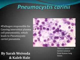
Pneumocystis carinii pneumonitis (PCP) is a common opportunistic disease that occurs almost exclusively in persons who have profound immunodeficiency.PCP was and still is the most common life-threatening opportunistic infection occurring in patients with HIV disease.
The taxonomy of P carinii has not been established. It is either a protozoan or a fungus. Recent studies show P carinii more closely resemble fungi than protozoa. rRNA sequences thymidylate synthase dihydrofolate reductase beta tubulin mitochondrial DNA chitin in the cell wall O E Eriksson has a treatise which places P carinii in a new family, Pneumocystidaceae, and in a new order, Pneumocystidales (Ascomycota).
The mode of replication of P carinii has not been established. However, the stages in its life cycle have been characterized. Sporozoites excyst through breaks in the cyst wall and then are termed trophozoites. The means by which the trophozoite form progresses to the cyst phase is not known.
The portal of entry for P carinii has not been firmly established; however, because with rare exceptions the organism has been found only in the lung, inhalation is a likely mode of transmission. Airborne transmission has been demonstrated in animals. In most individuals, the organism is dormant and sparsely dispersed in the lung, with no apparent host response (latent infection). In susceptible (immunocompromised) hosts, the organism occurs in massive numbers.
With rare exceptions, P carinni causes disease only when natural mechanisms of host defense are compromised. Pneumonitis tends to occur in patients with impaired cell-mediated immunity, and it is a major infection in patients with the acquired immune deficiency syndrome (AIDS). Severe protein-calorie malnutrition alone may provoke the disease. Immunosuppressive drugs used for cancer or organ transplantation render the individual susceptible to P carinii pneumonitis.
Pneumocystis carinii has been found in the lungs of rats, rabbits, mice, dogs, sheep, goats, ferrets, chimpanzees, guinea pigs, horses, and monkeys. The organism has been reported in lower animals and humans from all continents. Animal to animal transmission by the airborne route has been demonstrated. Because about 70 percent of healthy individuals may have humoral antibody to P carinii, subclinical infection must be highly prevalent.
Tachypnea and fever are consistent features of the pneumonitis, and diffuse bilateral alveolar disease can be observed by radiography. Diagnosis requires the identification of P carinii in pulmonary tissue or lower airway fluids. Such specimens may be obtained by lung biopsy, inducement of sputum, bronchoalveolar lavage, or needle aspiration of the lung. The Gomori, Giemsa, fluorescence-labelled antibody, or toluidine blue O stains may be used to identify the organism.
Genus/Species: Pneumocystis carinii • Image Type: Microscopic Morphology • Title:EM Image of Pneumocystis carinii • Disease(s): Pneumocystis pneumonia • Legend:An electron micrograph of P. carinii cyst from rat lung tissue. Pneumocystis carinii http://www.doctorfungus.org
Genus/Species: Pneumocystis carinii • Image Type: Microscopic Morphology • Title:Pneumocystis carinii-infected • Rat Lung Tissue • Disease(s): Pneumocystis pneumonia • Legend:An H&E stain of a rat lung infected with P. carinii. It does not show any organisms, but shows the proteinaceous exudate which results from Pneumocystis infection, and ultimately causes reduced gas exchange. Pneumocystis carinii http://www.doctorfungus.org
Genus/Species: Pneumocystis carinii • Image Type: Microscopic Morphology • Title:Pneumocystis carinii Silver Stain Disease(s): Pneumocystis pneumonia • Legend:A silver stain of P. carinii cysts from rat lung tissue showing the typical 'deflated ball' shape. Pneumocystis carinii http://www.doctorfungus.org
Genus/Species: Pneumocystis carinii • Image Type: Microscopic Morphology • Title:EM Image of Pneumocystis cariniiDisease(s): Pneumocystis pneumonia • Legend:An electron micrograph of a P. carinii troph from rat lung tissue, showing its binding • to a type I pneumocyte. Pneumocystis carinii http://www.doctorfungus.org
Four drugs currently available for therapy of P carinii pneumonitis are: • pentamidine isethionate • trimethoprim-sulfamethoxazole • atovaquone • trimetrevate Trimethoprim-sulfamethoxazole is preferred because of its low toxicity and greater efficacy.
Information obtained from: • UTMB Graduate School of Biomedical Sciences http://gsbs.utmb.edu • Dr Fungus http://www.doctorfungus.org • HIV Insite http://hivinsite.ucsf.edu