Download
1 / 12
400 likes | 2.27k Views
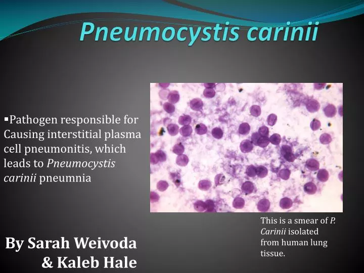
Pneumocystis carinii. Pathogen responsible for Causing interstitial plasma cell pneumonitis , which leads to Pneumocystis carinii pneumnia. By Sarah Weivoda & Kaleb Hale. This is a smear of P. Carinii isolated from human lung tissue. Taxonomic Considerations .

E N D
Pneumocystis carinii • Pathogen responsible for Causing interstitial plasma cell pneumonitis, which leads to Pneumocystiscariniipneumnia By Sarah Weivoda & Kaleb Hale This is a smear of P. Carinii isolated from human lung tissue.
Taxonomic Considerations • Currently considered to be a fungus -Based on nucleic acid analysis • Previously considered to be a protozoa Domain: Eukaryota Kingdom: Fungi Phylum: Ascomycota Class: Pneumocystidomycetes Order: Pneumocystidales Family: Pneumocystidaceae Genus: Pneumocystis Species: carinii
History of P. Carinii • First discovered in 1909 by Carlos Chagas • Found to be associated with clinical pneumonia shortly after World War 2 • Until the 1980’s it occurred very rarely and only caused pneumonia in people with congenital immunodeficiency's and patients immunocompromised by cancer. • From the 1980’s on the incidence of Pneumocystis Carinii associated pneumonia significantly increased due to the AIDS epidemic. • P. Carinii is now the leading cause of death by opportunistic infection in AIDS patients.
Infects a broad range of mammalian species including: -Humans -Mice -Rats -Cats -Dogs -Pigs Geographic range • Affects Humans and animals world wide -Most healthy children have been exposed to P. Carinii by age 3 to 4 -Primarily affects immunocompromised individuals
Definitive Host • Immunocompromised Humans and many other mammalians such as mice, rats, cats, dogs and pigs. P. Carinii has no known intermediate host or vector species
Pathogenesis • The specific mode of transmission is unknown but evidence suggest airborne transmission. • Causes disease by growing and filling the alveoli of the lungs
Clinical Signs Physical Symptoms Initial *Fever *Fatigue *Weight Loss Dyspnea Tachypnea Nonproductive cough Fevers Chills Sweats Progressive, profound fatigue Cyanosis around the mouth, hands, feet, or mucous membranes • Progressive • Extrapulmonary manifestations • Death
Cyst Laboratory Diagnosis Trophozoite
Morphology and Biology • P. Carinii goes through 3 morphological stages: 1. Trophozoite • pleomorphic in shape • Ranges from 1 to 5 µm in diameter • Has small filopodia 2. Precyst • Oval in shape • Has few filopodia • Has a cluster of mitochondria in its center 3. Cyst - Spherical in shape • Has a thick membrane made of chitin • Contains 8 intracystic bodies All morphological stages can be found within the lungs of the infected individual
Treatment • Drug of choice • Trimethoprim-sulfamethoxazole • Recommended others • Pentamidine • Trimethoprim plus daposone • Atovaquone • Primaquine plus clindamycin
References • http://dpd.cdc.gov/dpdx/HTML/Pneumocystis.htm • http://www.tulane.edu/~wiser/protozoology/notes/aids.html • http://www.aafp.org/afp/991015ap/1699.html • http://emedicine.medscape.com/article/225976-overview • http://www.nlm.nih.gov/medlineplus/ency/article/000671.htm • Roberts, L. Janovy, J. Foundations of Parasitology, 8th ed. New York: McGraw-Hill, 2009.