Download
1 / 63
970 likes | 2.58k Views
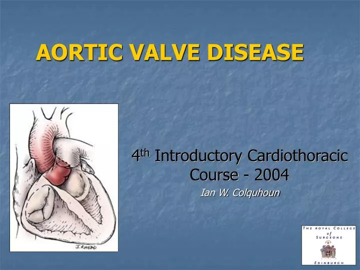
AORTIC VALVE DISEASE. 4 th Introductory Cardiothoracic Course - 2004 Ian W. Colquhoun. ANATOMY. Aortic Root - Anatomy. The AORTIC ROOT has four anatomic components:. The aortic annulus or aortoventricular junction. The leaflets. SINGLE FUNCTIONAL UNIT.

E N D
AORTIC VALVE DISEASE 4th Introductory Cardiothoracic Course - 2004 Ian W. Colquhoun
Aortic Root - Anatomy The AORTIC ROOT has four anatomic components: • The aortic annulus or aortoventricular junction • The leaflets SINGLE FUNCTIONAL UNIT • The aortic sinuses or sinuses of Valsalva • The sinotubular junction
Aortic Annulus (aortoventricular junction) N L R Sinotubular Junctinon Fibrous tissue Myocardium Anterior leaflet of the mitral valve Membranous septum Normal: 55% 45% Marfan/Bicuspid aortic valve: 65% 35% Histology: The aortic root is in fibrous continuity with the anterior leaflet of the mitral valve and the membranous septum; connective tissue (fibrous strands) unites the aortic root to the interventricular septum.
Valve Leaflets They are attached to the aortic root in a semilunar fashion The triangular space underneath the leaflet (trigone) is part of the left ventricle. The highest point of the trigone where the leaflets meet is called the commissure. The commissures are localised immediately below the Sinotubular Junction. The 2 trigones underneath the commissures of the noncoronary leaflet are fibrous structures, whereas the other underneath the commissure between the right and the left leaflets is mostly a muscular structure.
Sinuses of Valsalva The segment of the arterial wall of the aortic root delineated by a leaflet proximally and by the sinotubular junction distally is called the aortic sinus or sinus of Valsalva. • They are 3 elliptical inlets that have a very important role in the dynamics of circulation : • Guaranteeing coronary artery • perfusion during systole; • Creating eddies to close the • aortic leaflets during diastole
Sinotubular Junction It represents the terminal edge of the aortic root and it is constituted by the imaginary line that connects together the 3 commissures. Young adults AA>STJ Adults AA = STJ Elderly AA<STJ BASE = Aortic Annulus (AA)
Anatomic and Echocardiographic Relationship Between the Components of the Normal Aortic Root 120 degree - LAX Systole Annulus ST junction Tubular aorta Sinuses Diastole
Morphology • Calcific Aortic Stenosis Congenitally bicuspid or unicuspid, fused commissures, heavy calcification, age 40-60 1% Population • Rheumatic Aortic StenosisFibrous thickening, 3-cusp valve, mild calcification, commisural fusion, rheumatic fever history in ½ • Degenerative Aortic Stenosis Diffuse nodular calcification 3-cusp valve, no commissural fusion
Aortic Stenosis – Aetiology / Age AGE >70 AGE <70
Levels of stenosis (1) • SUPRA – VALVAR • WILLIAMS SYNDROME • Elfin-like facies • Hypervitaminosis D • Pulmonary stenoses – valvar & peripheral • Mesenteric artery stenosis • Thoracic aneurysm
Levels of stenosis (2) • SUB – VALVAR • DISCRETE FIBROMUSCULAR RING • 10% congenital AS • Presents <1year of age • 50% have other cardiac defects • HOCM • ANOMALOUS ATTACHMENT of MITRAL VALVE • AV canal • Parachute deformity of mitral – fused papillary muscles
Aortic Stenosis: Pathophysiology • Normal valve area = 2.5 – 3.5 cm2 • Gorlan Formula MILD AS >1.2cm2 MODERATE 1.0 – 1.2 cm2 SEVERE 0.8 – 1.2 cm2 CRITICAL <0.8cm2 • Severe AS & normal cardiao output = transvalvar gradient >50mmHg • Pressure overload • Concentric left ventricular hypertrophy increased wall stress, decreased ejection fraction reduced coronary reserve, subendocardial ischaemia • Increased LVEDP Higher preload required Left atrial hypertrophy, prominent “a” wave Loss of sinus rhythm – serious clinical deterioration • Eventual failure and dilatation of LV
AS: Clinical Picture Symptoms • Asymptomatic • Syncope • cerebral hypoperfusion vs. dysrhythmias • Angina • myocardial O2 supply/demand imbalance • Congestive Heart Failure • Sudden death
AS: Diagnosis CVS: • Loud harsh crescendo-decrescendo murmur radiating to neck • Possible diastolic murmur (AI murmur) • Split S2, possible S4 with atrial hypertrophy • Prominent presystolic apical impulse • Diminished carotid upstroke ECG: • LVH • LA hypertrophy • conduction abnormalities
AS: Diagnosis CXR • Left ventricular prominence with possible cardiomegaly • Calcifications at level of aortic valve Echocardiogram with Doppler Heart Catheterization • Coronary artery anatomy recommended for all pts • Visualization of aorta • Quantify gradient across valve • Calculate aortic valve area with Gorlan formula • Quantify aortic regurgitation • Assess mitral valve, regional wall motion disturbances, and left ventricular function
Natural History - Stenosis a) HAEMODYNAMICALLY SEVERE (symptomatic or asymptomatic) · Sudden death risk high· Immediate operation is indicated b) HAEMODYNAMICALLY MILD – MODERATE (asymptomatic) · 50% event free for 4 years· Operation is not urgent, but patients should be followed carefully as the disease advances rapidly c) HAEMODYNAMICALLY MILD – MODERATE (symptomatic) · One-third will die within 4 years· Prompt operation is indicated • Natural History Progression average of 0.1cm2 per year • Onset of Symptoms correlates with mortality risk
AR : Pathophysiology • Cusp Perforation • Cusp Prolapse • Restrictive Motion • Sinotubular Junction Dilatation • Annulus Dilatation • Annulo-aortic Ectasia
AR due to Abnormalities of the Leaflets Infective endocarditis Trauma Iatrogenic cause Cusp perforation
AR due to Abnormalities of the Leaflets • Excess of tissue • Disrupted commissure • Commissural malposition Cusp prolapse
AR due to Abnormalities of the Leaflets Fibrous thickening Restrictive motion
AR - Abnormalities of the Aortic Wall Sinotubular junction dilatation Dilatation of the sinotubular junction displaces the commissures outward and prevents the aortic leaflets from coapting, with resulting central aortic insufficiency
AR - Abnormalities of the Aortic Wall • Marfan syndrome • Connective tissue diseases • Ventricular dilatation • Chronic hypertension Annulardilatation
AI due to Abnormalities of the Aortic Wall Aortic root aneurysm: ST junction dilatation + Sinuses of Valsalva aneurysm
Aortic Regurgitation (1) • CONGENITAL • Bicuspid valve • Supra-valvar stenosis • Supra-cristal VSD and right coronary prolapse • Sinus of Valsalva aneurysm
Aortic Regurgitation (2) ACQUIRED • VALVE - Cusp prolapse or cicatricial shortening of cusps with rolled edges • Rheumatic fever • Infective endocarditis • Rheumatoid disease • SLE • Hurler’s syndrome • AORTIC ROOT - Dilation of sinus aorta, failure of coaptation of cusps • Dissection • Syphilis • Cystic medial necrosis e.g Marfans – annulo-aortic ectasia • Arthritides with aortitis e.g. Ankylosing spondylitis • Hypertension • Trauma
Aortic Valve Insufficiency It may be due to alteration of the valve, the ascending aorta or both. Etiology: • Idiopathic degenerative disease • Aortic dissection • Calcific aortic disease • Myxomatous degeneration • Rheumatic or postinflammatory disease • Bicuspid aortic valve • Trauma • Infective endocarditis • Idiopathic dilatation of the aortic root • VSD of the membranous septum • Systemic diseases(Whipple • disease; Crohn’s disease;) • Aortitis(Syphilis, viral • syndromes; giant cell • arteritis;Takayasu disease; • Chronic systemic hypertension • Connective tissue disorders • (Marfan’s syndrome; Reiter’s • disease; Ehlers-Danlos • syndrome; Osteogenesis • imperfecta; Rheumatoid • arthritis syndrome; Ankylosing • Spondylitis; SLE;)
Aortic Valve Insufficiency • Annulus • Annulo-aortic ectasia • Leaflets • Prolapse • Degeneration • Infectious disease Valve Sparing Acute Chronic • Sinuses of Valsalva • Dilatation of one or more sinus • Ascending aorta dissection • Sinotubular Junction • Global dilatation (including ascending aorta)
AR - Diagnosis Eponyms associate with AR • Austin-Flint murmur – vibrations of anterior mitral leaflet • Duroziez’s sign – ‘to and fro’ femoral artery murmur • Quincke’s pulse – capillary pulsation in finger tips • Traube’s sign – ‘pistol shot’ sound at femoral artery • De Musset’s sign – head bobbing
Natural History - Incompetence a) Latent period to cardiac decompensation is long · Sudden death is not common· Once deterioration begins, the LV fails rapidly b) Symptomatic patient with CHF, angina, syncope · Prompt operation is indicated c) Asymptomatic patient ·Follow carefully for LV enlargement or decreased LV function by ECHO or MUGA· Operate at an appropriate time
Associated Coronary Artery Disease · Treat significant coronary artery disease at the time of surgery even if asymptomatic · CABG reduces risk of AVR and improves long-term survival · Coronary angiography is indicated in all patients older than 45 years who will be having AVR
ACC/AHA Guidelines for the Management of Patients With Valvular Heart Disease. Executive Summary. A report of the American College of Cardiology/American Heart Association Task Force on Practice Guidelines (Committee on Management of Patients With Valvular Heart Disease). Bonow RO, Carabello B, de Leon AC, Edmunds LH Jr, Fedderly BJ, Freed MD, Gaasch WH, McKay CR, Nishimura RA, O'Gara PT, O'Rourke RA, Rahimtoola SH, Ritchie JL, Cheitlin MD, Eagle KA, Gardner TJ, Garson A Jr, Gibbons RJ, Russell RO, Ryan TJ, Smith SC Jr. J Heart Valve Dis. 1998 Nov;7(6):672-707
ASYMPTOMATIC Mild – moderate stenosis Medical follow up Regular ECHO Avoid strenuous exercise Endocarditis prophylaxis ? Role for statins Progress ~0.1cm2 per year SYMPTOMATIC AVR Angina, syncope, failure Moderate AS + CAD Reduced BP on exercise Severe AS & reduced LV function MANAGEMENT - AS
MEDICAL Calcium channel blocker Regular ECHO Avoid isometric exercise Endocarditis prophylaxis Monitor for symptoms Protracted course SYMPTOMATIC AVR Symptom onset Asymptomatic if: LVESD > 50-55mm LVEDD > 70-75mm LVEF < 55% MANAGEMENT - AR
Age and AVR · Advanced age most common predictor of survival and cardiac events· AVR very effective treatment even in patients over age 70 or 80· Even the best patients over age 80 have reduced reserve
Trends in choice of prosthesis · Age less than 55 years - Aortic allograft or pulmonary autograft · Age between 55-75 years - Mechanical prosthesis · Age greater than 75 years - Porcine heterograft, stented or stentless · Allografts and autografts enlarge the orifice by about 2 mm, porcine heterografts reduce valve size by about 2 mm, and mechanical valves reduce valve size by about 5-8 mm
Operative Principles • Restore unimpeded flow through aortic annulus / left ventricular outflow tract • Remove stenosis / regurgitation • Match valve size to patient • Repair / replace abnormal aorta ± reimplantation of coronary arteries • Concomitant pathology addressed: CABG Mitral valve
Pitfalls • Inadequate decalcification • Too vigorous decalcification • Heart block from suture • Coronary ostial occlusion from sewing ring • Debris lodging in Left coronary ostium
Operative Results • Increased risk if • Emergency • NYHA Class III – IV • >65 years old • Severe AS AVA<0.7 or AVG>70, LVEDP>20 • Impaired LV systolic function • Need for other procedure (CABG) • Renal dysfunction • Small BSA • Redo operation • Post op complications: • Operative mortality: ~ 5% • Complete heart block • Ischemic heart disease (6 months from coronary ostial stenosis) • CVA 3 – 5%
Long term survival • 5 year survival • Normal LV function: 96% • Overall • 80% @ 5 years • 60% @ 10 years • Depressed LV function: 63% • Valve related morbidity responsible for >50% late deaths • Bioprosthetic valve - Reoperation: • 25% @ 10 years • 65% @ 15 years