Download
1 / 23
320 likes | 898 Views
Mitral Valve Disease. Prof JD Marx UFS January 2006. Anatomy. Mitral Stenosis. Aetiology. Almost always rheumatic Heavy calcification in elderly Congenital MS in infants. Pathophysiology. Mitral valve orifice diminished by progressive fibrosis calcification valve leaflets

E N D
Mitral Valve Disease Prof JD Marx UFS January 2006
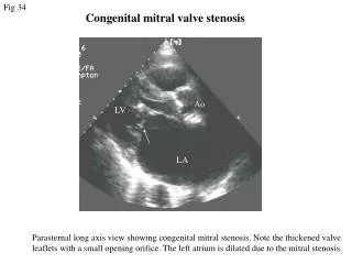
Mitral Stenosis Aetiology • Almost always rheumatic • Heavy calcification in elderly • Congenital MS in infants
Pathophysiology • Mitral valve orifice diminished by • progressive fibrosis • calcification valve leaflets • fusion cusps subvalvular apparatus
Mitral Valve Orifice • Normal 5 cm² • Moderately severe 2 cm² or less • severe 1 cm² or less • Pulmonary venous congestion • LA dilatation and hypertrophy • LA contraction important LV filling • Diastolic filling period important • Pulmonary hypertension • Atrial fibrillation • Thrombus formation in LA Bloodflow LA to LV restricted
Symptoms • Breathlessness (pulmonary congestion) • Fatigue (low cardiac output) • Oedema, ascites (right heart failure) • Palpitation (atrial fibrillation) • Haemoptysis (pulmonary congestion, pulmonary embolism) • Cough (pulmonary congestion) • Chest pain (pulmonary hypertension) • Symptoms of thromboembolic complications (e.g. stroke, ischaemic limb)
Signs • Atrial fibrillation • Mitral facies • Auscultation • Loud first heart sound. Opening snap • Mid-diastolic murmur • Signs of raised pulmonary capillary pressure • Crepitations, pulmonary oedema, effusions • Signs of pulmonary hypertension • RV heave, loud P2
Investigations • ECG • Left atrial hypertrophy (if not in AF) • Right ventricular hypertrophy • Chest Radiograph • Enlarged left atrium • Signs of pulmonary venous congestion • Echo • Thickened immobile cusps • Reduced valve area • Reduced rate of diastolic filling of LV • Doppler • Pressure gradient across mitral valve • Pulmonary artery pressure • Cardiac catheterisation • Pressure gradient between LA (or pulmonary wedge) and LV
Management • Medical management • Patients with minor symptoms • Anti coagulants eg Warfarin • Diuretics for pulmonary congestion • Rate and rhythm control digoxin, -blockers etc • A/B prophylaxis IE
Surgical procedures • Mechanical disease • Consider - patient symptomatic • - pulmonary hypertension • - atrial fibrillation • - MVA 1 cm² or less • Mitral balloon valvuloplasty • Open mitral replacement
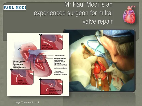
Mitral Regurgitation Aetiology • Chronic rheumatic endocarditis • Infective endocarditis • Mitral valve prolaps and myxomatous degeneration • Mitral valve ring dilatation eg dilating cardiomyopathy • Papillary muscle necrosis / ischaemia
Pathophysiology • Chronic mitral regurgitation • Gradual dilatation LA • Little increase in pressure • LV dilates slowly • Late rise in diastolic and LA pressure • Acute mitral regurgitation • Rapid rise in LA pressure (compliance)
Clinical Features Symptoms • Dyspnoea (pulmonary venous congestion) • Fatigue (low cardiac output) • Palpitation (AF, increased stroke volume) • Oedema, ascites (right heart failure)
Signs • Atrial fibrillation / flutter • Cardiomegaly – displaced hyperdynamic apex beat • Apical pansystolic murmur ± thrill • Soft S1, apical S3 • Signs of pulmonary venous congestion (crepitations, • pulmonary oedema, effusions) • Signs of pulmonary hypertension and right heart failure
Investigations • ECG • Left atrial hypertrophy (if not in AF) • Left ventricular hypertrophy • Chest Radiograph • Enlarged left atrium • Enlarged left ventricle • Pulmonary venous congestion • Pulmonary oedema (if acute) • Echo • Dilated LA, LV • Dynamic LV (unless myocardial dysfunction predominates) • Structural abnormalities of mitral valve (e.g. prolapse) • Doppler • Detects and quantifies regurgitation • Cardiac catheterisation • Dilated LA, dilated LV, mitral regurgitation • Pulmonary hypertension • Coexisting coronary artery disease
Management • Medical management • Mild to moderate MR • Diuretics • Vasodilators eg ACE Inhibitors • Digoxin if AF • Anti coagulants if AF • A/B prophylaxis for IE • Surgical management • Patient more symptomatic • Evidence deteriorating LV function and LV dilatation • Mitral valve repair • Mitral valve replacement