Download
1 / 42
420 likes | 498 Views
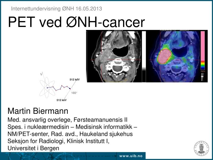
n. 512 keV. 180°. 512 keV. Internettundervisning ØNH 16.05.2013. PET ved ØNH-cancer.

E N D
n 512 keV 180° 512 keV Internettundervisning ØNH 16.05.2013 PET ved ØNH-cancer Martin BiermannMed. ansvarlig overlege, Førsteamanuensis IISpes. i nukleærmedisin – Medisinsk informatikk – NM/PET-senter, Rad. avd., Haukeland sjukehusSeksjon for Radiologi, Klinisk Institutt I,Universitet i Bergen
PET ved hode/hals cancer FDG-PET ved hode/hals-cancer • Generelt om PET • FDG-PET ved hode/hals-cancer: • Primærstaging • Doseplanlegging ved stråleterapi • Multimodal utredning ved ca. thyreoideaeved PET-senteret i Bergen
PET ved hode/hals cancer Tromsø Mobil PET/CT 2011: 222 2012: 234 PET/CT i Norge 2011: 4119 2012: 5413 Oslo Syklotron + 4 PET/CT 2011 2012 Rikshospitalet 1338 1511 Radiumhospitalet 1034 1268 Ullevål 509 957 Aleris 146 156 SUM 3027 3892 . Bergen Syklotron + 1 PET/CT 2011: 870 2012: 1287 Rune Sundset, UNN
PET ved hode/hals cancer Røntgen versus nukleærmedisin/PET gamma-kamera/PET-kamera røntgenrør pasient radioaktivtlegemiddel film Påvise endringer i metabolismefør anatomi er påvirket anatomisk bilde metabolisk bilde
n 180° PET ved hode/hals cancer Positronstråling (b+) Positroner = anti-elektroner • positron bremses i vevet i løpet av 1 – 2 mm • kollisjon med elektron:annihilasjonsstråling E = mc2 – 2 fotoner i motsatt retning
PET ved hode/hals cancer Gammakamera v. PET • PET (versus NM): • oppløsning 4 mm (vs. 1 - 2 cm) • alltid 3-dimensjonal kollimator koinsidens-deteksjon Gammakamera PET
PET CT PET ved hode/hals cancer PET-CT
GLUT FDG-P FDG-P Gluc FDG-P Gluc Gluc Gluc Gluc Gluc PET ved hode/hals cancer F-18-fluorodeoksyglukose (FDG) • Glukose merket med radioaktivt fluor-18 (F-18) • Forbrenning av glukose i kroppen er universell: • normalt: hjerne, hjerte, muskulatur • patologisk: mange solide svulster kreftdiagnostikk !! leukocytter betennelsesscintigrafi ”trapping” Hexokinase FDG Gluc-6-P
PET ved hode/hals cancer Indikasjoner for F18-FDG: Onkologi • Hode/halskreft1,2 • Thyreoidea1 • Øsofagus1,2 • Lungecancer (non-small-cell) 1,2 • Kolon/rektum1,2 • Lymfom1,2 • Melanom1,2 • flere andre indikasjoner: seminom, osteosarkom… 1Reske (2001) Eur J Nucl Med 28:1707-232http://www.cms.hhs.gov Fletcher (2008) J Nucl Med 49:480-508
PET ved hode/hals cancer Hode/halskreft • Prospektiv studie:n = 60 pas. • Resultater: 117 metastaser i 1284 lymfeknuter Sens. Spes.PET 90 % 94 %CT 82 % 85 %MR 85 % 94 %US 72 % Adams (1998) Eur J Nucl Med 25:1255-60
PET ved hode/hals cancer FDG-PET (CT) for hode/hals-kreft 2 metaanalyser Analyse per nodallevel, gullstandard = histologi PET-CT økerspesifisitet over PET Kyzas (2008) J Natl Cancer Inst 100:712-20Yongkui (2013) Surg Oncol (in press)
PET ved hode/hals cancer FDG-PET (CT) for hode/hals-kreft 76 konsekutive pas. planlagt for neck dissection FDG-PET best ved N+Cave: nekrotiske & inflammatoriske LN Analyse N0/N+ (per pasient), gullstandard = histologi FNB mest spesifikk Stoeckli (2012) Head & Neck 34:469-76
PET ved hode/hals cancer FDG-opptak og lokal kontroll • 50 pas. med ny plateepitel-CA i hode/hals Høyt FDG-opptakdårlig prognose Torizuka (2009) AJR Am J Roentgenol 192:W156-60
PET ved hode/hals cancer Case 1 f 75 yrs. SCC retromolar L op. Anatomien fremstilles KUN ved KM-CT KM-CT LD-CT
PET ved hode/hals cancer Case 1 f 75 yrs. SCC retromolar L OP 2002 cancer! LK-met. LIIb M. pterygoideus medialis ! IKKE cancer!
PET ved hode/hals cancer Tumor volume squamous epithelial cancer • Methods: • total laryngectomy in 9 pts. • CT, MR, FDG-PET with mask • OP preparation kryofixated en bloc • All methods overestimated tumor volume (TV): • PET most accurate • 20 % of TV only seen with PET • PET essential for radiotherapy planning Daisne (2004) Radiology 233:93-100
PET ved hode/hals cancer Dose planning for RTx with PET-CT Haukeland University Hospital 14 Apr 2010
PET ved hode/hals cancer Case 2 m 78 yrs. SCC epipharynx T2 N2 M0 FDG-uptake in primary tumor + 3 lymph nodes
PET ved hode/hals cancer Case 2 m 78 yrs. SCC epipharynx T2 N2 M0 Need for a multimodal diagnostic viewing system on top of the dose planning system ! Read each modality on its own !
PET ved hode/hals cancer Case 2 m 78 yrs. SCC epipharynx T2 N2 M0
PET ved hode/hals cancer Treatment plan for oropharyngeal cancer Diagnosis chemotherapy IMRT 6 0 weeks PET-CT PET-CT
PET ved hode/hals cancer Case 3 m 51 yrs. SCC vallecula T2 N2c M0 renal failure:no CE-CT Metabolic response: FDG-uptake in the GTV pre-chemo PET-CT essential
PET ved hode/hals cancer IMRT plan m 51 yrs. SCC vallecula T2 N2c M0
PET ved hode/hals cancer FDG-PET for radiation therapy planning • Firmly established clinical routine for head & neck and pelvic cancers at Haukeland since 2010 even before own FDG-production • Clinical workflow:close collaboration between radiologist, nuclear physician, radiooncologist, physicist, technicians • Challenges in multimodal imaging: • One MUST view all modalities on their own and in geometric correlation (fusion) to each others: Dedicated multimodal diagnostic viewing systemin addition to the radiation therapy planning system • Quality assurance with phantoms
Medviz Seminar 12 Apr 2013 Multimodal imaging for suspected recurrent differentiated thyroid cancer (DTC) FDG-PET-CT, I-131 SPECT-CT, ultrasound (US) andUS-guided fine needlebiopsy (FNB) The incremental value of multimodality imaging oversingle-modality US & PET M. Biermann,1,4 J. Kråkenes,1,4 Haugland HK,2L. Akslen,2,4 J. E. Varhaug,3,4 M. Brauckhoff3,4 1PET-centre, Dept. of. Radiology, 2Dept. of Pathology3Dept. of Endocrine Surgery, Haukeland University Hospital4University of Bergen, Bergen, Norway
PET ved hode/hals cancer Material and Methods • n = 51 patients with newly suspected recurrencefrom 6/2009 – 12/2011 at Haukeland University Hospital in Bergen/Norway • Ultrasound + fine needle biopsy (FNB) before PET • I-131-SPECT-CT (≥ 3 GBq)* • F-18-FDG-PET* incl.contrast-enhanced CT (CE-CT) of the neck • Serum-hTg* • Ultrasound + FNB after PET * under TSH stimulation Biermann (2003) Eur J Nucl Med Mol Imaging 30:160-3 van Tol (2002) Thyroid 12:381-7
PET ved hode/hals cancer Results: Multimodal imaging (MMI) • 51 patients: accuracy 92 % • 63 neck & mediastinal lesions in 24 patients • 43 malignant: 5 local recurrences, 5 mediastinal • 20 benign • histology in 39, FNB in 11, imaging follow up in 13 • Distant metastases in 10 pts. (9 lung, 3 bone, 1 adrenal) accuracy 86 %
PET ved hode/hals cancer Neck lesions (n = 57) • Pre-PET ultrasound: accuracy 49 % • FDG & I-131 uptake: accuracy 80 % • Post-PET ultrasound: accuracy 88 %
PET ved hode/hals cancer Therapeutic consequence Pre-PET intendedTx vs. post-PET actualTxin the true positive patients/ all patients (= 51): • 10 noTx activelocalTx (8 OP, 1 laser, 1 RTx)(lesionoverlooked in pre-PET US in 13 pts.*) • 3 surgery extendedsurgery in other region(lesionoverlooked in pre-PET US in 3 pts.) • 1 surgery noTx(false pos. pre-PET US) • 18 surgery as intended S Treatmentchanges in 14/51 pts. = 28 % *3 patients "too old" for surgery
PET ved hode/hals cancer Case 1 f. 50 yrs. Pap. TC stim. hTg 1490 ng/ml SUVmax 10 FDG-pos. paratracheal recurrence !? F-18-FDG-PET I-131-SPECT-CT (3 GBq)
PET ved hode/hals cancer Case 1 f. 50 yrs. Pap. TC stim. hTg 1490 ng/ml FNB:paratracheal recurrence Therapy:endotracheal laser ablation 400 xL. Helgeland HUS
PET ved hode/hals cancer Case 2 f. 55 yrs. PTC OP 2006 ↑↑Tg-AB hTg 0 400 xL. Helgeland HUS FNB:Papillary thyroid cancer!
PET ved hode/hals cancer Case 2 FDG-PET-CT f. 55 yrs. PTC OP 2006 LN-metastasis paratracheal rec. Surgery (K1a, 2, 3) Haukeland:All 3 foci confirmedIntact recurrent laryngeal nerves US + FNB: 1 LN-metastasisPET: 2 extra tumour lesions! MIP LN-metastasis
PET ved hode/hals cancer Case 3 f. 43 yrs. pap. TC stim. hTg < 0.2 ng/ml I-131 I-131 positive metastasis F-18-FDG SUVmax 2 CE-CT
PET ved hode/hals cancer Case 3 f. 43 yrs. pap. TC stim. hTg < 0.2 ng/ml Cranial FDG-pos. LN !de-differentiated metastasis? FDG SUVmax 2 I-131 FDG SUVmax 4 CE-CT CE-CT
PET ved hode/hals cancer Case 3 f. 43 yrs. pap. TC stim. hTg < 0.2 ng/ml 400 xHK Haugland HUS FDG SUVmax 4 US guided FNB: Tg < 0.2inflammatory LN ! OP 1/2011: 1 LN metastasis (I-131 pos.)22 tumor-free LN CE-CT
PET ved hode/hals cancer Case 4 f. 56 yrs. pap. TC stim. hTg 5 ng/ml Microcalcifications!Metastasis? OP 1/2011: 1 LN metastasis (I-131 pos.)22 tumor-free LN Hyperperfusion:Metastasis !? Doppler ultrasound
PET ved hode/hals cancer Case 4 f. 56 yrs. pap. TC stim. hTg 5 ng/ml FDG & I-131 negative! FDG SUVmax < 1 OP 1/2011: 1 LN metastasis (I-131 pos.)22 tumor-free LN Doppler ultrasound FDG-PET + CE-CT
PET ved hode/hals cancer Case 4 f. 56 yrs. pap. TC stim. hTg 5 ng/ml 200 xHK Haugland HUS OP 5/2011: 1 LN metastasis 17 tumor-free LN OP 1/2011: 1 LN metastasis (I-131 pos.)22 tumor-free LN Fine needle biopsy:thyroid epithelium hTg 800 ng/ml in washout FDG & I-131 neg. metastasis ! Doppler ultrasound
PET ved hode/hals cancer FDG-PET for residiv ved ca. thyreoideae • Multimodal billeddiagnostikk positiv hos 65 %Endret terapi hos 27 % (14/51) av pasienter Ikke operer uten PET! • Kun 80 % accuracy ved FDG-PET: • Submandibulære lymfeknuter: FNB ! • Pulmonale infiltrater, thymus • Fysiologisk/reaktivt opptak i muskulatur Post-PET ultralyd + finnålsbiopsi ! • Præ-PET ultralyd + FNB viktig for optimal indikasjon for FDG-PET
PET ved hode/hals cancer FDG-PET for hode/hals-cancer • Mest nytte ved N1 hals – mangler sensitivitet for å kunne «bevise» en N0-situasjon • Essensielt for stråleterapi-planlegging ved epipharynx-cancer:PET-CT før og etter neoadjuvant kjemoterapi • Cave:Falsk negativ FDG-opptak ved nekroserFalsk positiv ved inflammatoriske LKFDG-opptak enda mer variabel ved ca. thyr. • PET-CT best med koregistrert KM-CT hals:low dose CT = low-information CT!
PET ved hode/hals cancer Bergen PET-senter Fremtiden er multimodal !