Download
1 / 60
600 likes | 607 Views
Circulation: The Cardiovascular and Lymphatic Systems. Cardiovascular System. Cardiovascular system Consists of heart and blood vessels Encompasses blood circulation Delivers oxygen and nutrients to cells Carries away waste products Lymphatic system

E N D
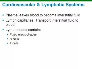
Cardiovascular System • Cardiovascular system • Consists of heart and blood vessels • Encompasses blood circulation • Delivers oxygen and nutrients to cells • Carries away waste products • Lymphatic system • Drains fluid and proteins from tissues, returns them to bloodstream
The Heart • Located between lungs • Myocardium = thick muscle layer • Endocardium = inside lining • Epicardium = Outside lining • Pericardium = surrounding fibrous sac
Location of Heart • The heart is encased in and separated from the walls of the pericardial cavity by three linings: the epicardium, which forms the outer part of the heart; the pericardial sac and the pericardium • The heart fits tightly inside the pericardial cavity, a subcavity of the thoracic cavityu. The pericardial cavity is lined with a serous ( thin) membrane called the pericardium, and the heart is within yet a second lining called the pericardial sac. This sac contains about half an ounce of fluid, which lies between it and the heart’s outer lining
The structure of the heart • The membrane forming the outer lining of the heart is called the epicardium • Immediately beneath the epicardium is the myocardium, comprising the muscles, blood vessels and nerve tissue that make up the bulk of the heart • The heart’s inner surface is called the endocardium
Heart (cont’d) • Atrium • Upper receiving chambers • Atria are separated by interatrial septum (plural septa) • Ventricle • Lower pumping chambers • Pulmonary circuit (right side to lungs) • Systemic circuit (left side to rest of body) • Ventricles are separated by interventricular septum • Also, each atrium is divided from each ventricle by an atrioventricular septum, which contains various valves
Heart valves • The right atrioventricular valve, also sometimes called the tricuspid valve, leads from the right atrium into the right ventricle • The pulmonary semilunar valve connects the right ventricle to the lungs, which also connect to the left ventricle through the left atrium by way of the left atrioventricular valve, also sometimes called the bicuspid ot mitral valve • The aortic semilunar valve leads out of the left ventricle
Blood Flow Through the Heart • When blood comes back to the heart after having delivered oxygen and other nutrients to the body’s cells, it needs to be replenished before going out again. It re-enters the heart at the atrium • Right atrium receives blood from body • Enters right ventricle and is pumped to lungs • Oxygenated blood returns to left atrium • Enters left ventricle and is pumped to rest of body • One-way valves force blood flow forward • Heart sounds produced when valves close
The Heartbeat • Systole = contraction (emptying the heart) • Diastole = relaxation ( refilling chambers) • Heart beats start with both atria contracting • Immediately thereafter both Ventricles contract • Wave of increased pressure in the vessels following ventricular contraction is pulse • Contractions are stimulated by electrical impulse Blood Pressure • Force of blood exerted against wall of blood vessel • Measured by sphygmomanometer • Measured as both systolic and diastolic, such as 120/80
Pacemaker and conduction system of the heart • Contractions are stimulated by a built-in system that regularly transmits electrical impulses through the heart • They include the sinoatrial (SA) node, called the pacemaker because it sets the rate of the heart beat • The atriopventricular (AV) node, the AV bundle (bundle of His) the left and right bundle branches and Purkinje fibers
Clinical Aspects of the Circulatory System • Atherosclerosis • Accumulation of fatty deposits within artery • Risk factors: • High levels of lipoproteins (especially LDL’s) • Smoking • High blood pressure • Poor diet • Inactivity • Stress • Family history
Thrombosis and Embolism • Definitions: • Thrombosis = formation of blood clot • Thrombus = blood clot • Embolism = blockage of blood vessel • Embolus = blockage mass • Blockage is usually blood clot • Blockage can also be air, fat, bacteria, or other solid materials • Stroke = blockage in a cerebral vessel
Aneurysm • Weakened arterial wall ballooning out • Caused by: • Atherosclerosis • Malformation • Injury
Heart Diseases • Coronary artery disease • Results from atherosclerosis • Early sign is angina pectoris (chest pain) • Diagnosed by: • ECG • Stress tests • Coronary angiography • Echocardiography • Treatments: • Control of exercise, administration of nitroglycerin • Angioplasty (PTCA) • Bypass (CABG)
Heart Diseases (con’t) • Myocardial infarction = heart attack • Symptoms: • Epigastric pain • Pain extending to jaw, arms • Pallor (turns pale) • Diaphoresis • Nausea • Dyspnea (difficulty breathing) • May also be burning sensation similar to heartburn
Heart Diseases (cont’d) • Arrhythmia • Irregularity of heart rhythm • Bradycardia = slower than average • Tachycardia = faster than average • Fibrillation = extremely rapid, ineffective • Controlled on Long term withpacemaker
Heart Diseases (cont’d) • Heart failure • Heart fails to empty effectively, leading to edema • Congenital heart disease • Birth defects • Most can be corrected surgically • Rheumatic heart disease • Streptococcus infection damaging heart valves
The Vascular System • Arteries and arterioles • Carry blood away from heart • Vasoconstriction and vasodilatation • Become smaller as they go away from the heart? • Capillaries • Smallest vessels • Where exchange between blood and tissues happens • Veins and venules • Carry blood back to heart
Conducting arteries • Sometimes called elastic arteries and can have an inside diameter as great as an inch • The aorta is an example of a conducting artery • The pulmonary artery and aortic trunk are examples of conducting arteries which move blood away from the heart • Three major conducting arteries branch from aortic arch. They are the brachiocephalic trunk, the left common carotid artery and the left subclavian artery • Both the right subclavian artery and the right common carotid artery attach to the brachiocephalic trunk
Medium size arteries, ( also called muscular arteries because they contain a lot of muscle tissue) typically have an inside diameter of about a sixth of an inch (eg external carotid artery) • Arterioles are the smallest arteries, with an average inside diameter of 0.0018 of an inch or about 1/100 the size of a medium size artery • Arteries and arterioles connect to the capillaries which can be as tiny as one blood cell ( or about ¼ the size of an arteriole in diameter)
Veins • Veins carry blood back to the heart. They follow the same path as the arteries ( with blood flowing in the reverse direction) • Also, like arteries, they vary in diameter, becoming larger as they approach the heart because of the increasing volumes of blood they must carry • The vein counterparts of the conducting arteries are the superior vena cava and the inferior vena cava. Together they are known as the venae cavae • Medium veins and venules are the counterparts of the muscular arteries and arterioles • Superior vena cava drains blood from upper body, including head neck shoulders and arms • Inferior vena cava receives blood from the lower body, the dividing line being the diaphragm
Disorders of the Veins • Varicose veins • Breakdown in valves with chronic dilatation • Contributing factors: • Heredity • Obesity • Prolonged standing • Pregnancy
Disorders of the Veins (con’t) • Phlebitis = inflammation of veins • Causes: • Infection • Injury • Poor circulation • Valve damage • Can result in thrombophlebitis (blood clot) • Most damaging if occurring deep
Hypertension • Commonly known as high blood pressure • Contributing factor in many conditions • Defined as systolic > 140, diastolic > 90 • Causes left ventricle to enlarge • First defense: diet and life habits
Blood Plasma • 90% water • Rest contains: • Nutrients • Electrolytes (dissolved salts) • Gases • Albumin (protein) • Clotting factors • Antibodies • Wastes • Enzymes • Hormones • Relative acidity (pH) steady at 7.4
Blood Cells • Produced in red bone marrow • Three kinds: • Red = erythrocytes • White = leukocytes • Platelets = thrombocytes
Erythrocytes • Main function: carry oxygen to cells • Most numerous of blood cells • Short lifespan (120 days) requires constant replacement • Production regulated by erythropoietin (hormone made in kidneys) Leukocytes Protect against foreign substances • Five different types: • Neutrophils • Eosinophils • Basophils • Lymphocytes • Monocytes
Leukocytes • Phagocytes are often the first immune system cells on the scene when injury occurs. They prevent infection by cleaning away pathogens and debris • Phagocytes are two types: microphages and macrophages • The average human body contains one trillion lymphocytes which include NK cells, T cells and B cells • T cells make up about 80% of the total number of lymphocytes • T lymphocytes depend on the thymus for their activation • T lymphocytes are prompted by a specific antigen; a substance that induces sensitivity
Leukocytes • Antigens also stimulate the immune system to generate antibodies, which can produce immunity from future attacks by the same type of antigen • Unlike NK cells which roam the body looking for intruders, T cells attack only when they recognize a specific antigen, and then only after receiving instructions from special T cells that distinguish between good and bad antigens • B lymphocytes are derived from bone marrow. Like NK cells, they roam the body looking for intruders • But unlike NK cells, they stop in lymph tissue to seek out foreign antigens • However they do not attack until the special T cells instruct them to do so
Platelets • Important for hemostasis • Most active during coagulation • Stick together to plug injury site • Interact with clotting factors in plasma to make wound-sealing clot • Convert fibrinogen to threads of fibrin • Threads of fibrin trap blood cells and plasma to make clot
Blood Types • Determined by genetically inherited proteins • Most familiar groups are ABO and Rh • Important to match for blood transfusions • Compatible types determined by cross-matching
The Immune System • Launches specific attacks on disease organisms • Involves components of lymphatic system and blood • Immune system response from T cells or B cells • T cells mature in thymus gland • B cells mature in lymphoid gland • Passive immunity: Transfer of antibodies • Naturally (mother’s milk) • Artificially (immune serum) • Active immunity: Individual’s own response to disease organism • Natural contact • Vaccine
Clinical Aspects: Blood • Anemia • Decrease in hemoglobin in blood • Can result from: • Too few red blood cells • Cells are too small • Too little hemoglobin • Key tests involve blood counts • Symptoms include fatigue, shortness of breath • Aplastic anemia = destruction of bone marrow • Nutritional anemia (includes pernicious anemia) = deficiency of vitamin B12 • Sideroblastic anemia = body doesn’t use iron properly • Hemorrhagic anemia = results from blood loss
Types of Anemia (con’t) • Thalassemia (includes Cooley anemia) • Hereditary disease causing rupture of red cells • Affects production of hemoglobin • Sickle cell anemia • Mutation alters hemoglobin molecule • Deformed cells block blood vessels and prevent tissues from receiving oxygen