Download
1 / 41
420 likes | 984 Views
The Lymphatic and Immune Systems. Chapter 20. The Lymphatic and Immune Systems. Main structures of the lymphatic system Lymphatic vessels Main components of the immune system Lymphocytes Lymphoid tissue Lymphoid organs. The Lymphatic System.

E N D
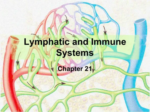
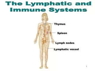
The Lymphatic and Immune Systems Chapter 20
The Lymphatic and Immune Systems • Main structures of the lymphatic system • Lymphatic vessels • Main components of the immune system • Lymphocytes • Lymphoid tissue • Lymphoid organs
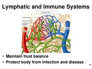
The Lymphatic System • Lymphatic vessels collect tissue fluid from loose connective tissue • Carry fluid to great veins in the neck • Fluid flows only toward the heart Figure 20.1
Functions of Lymphatic Vessels • Collect excess tissue fluid and blood proteins • Return tissue fluid and blood proteins to bloodstream
Orders of Lymphatic Vessels • Lymph capillaries -smallest lymph vessels • first to receive lymph • Lymphatic collecting vessels • collect from lymph capillaries
Orders of Lymphatic Vessels • Lymph nodes • scattered along collecting vessels • Lymph trunks • collect lymph from collecting vessels • Lymph ducts • empty into veins of the neck
Lymphatic Capillaries • Located near blood capillaries • Receive tissue fluid from CT - increased volume of tissue fluid - minivalve flaps open and allow fluid to enter • Highly permeability allows entrance of • tissue fluid • bacteria, viruses, and cancer cells
Lymphatic Capillaries • Lacteals – specialized lymphatic capillaries - located in the villi of the small intestines - receive digested fats - fatty lymph – chyle
Lymphatic Capillaries Figure 20.2a, b
Lymphatic Collecting Vessels • Accompany blood vessels • Composed of the same three tunics as BVs • Contain more valves than veins do • helps direct the flow of blood • Lymph propelled by • bulging of skeletal muscles • pulsing of nearby arteries • tunica media of the lymph vessels
Lymph Nodes • Cleanse the lymph of pathogens • Human body contains around 500 • Lymph nodes are organized in clusters
Lymph Nodes Figure 20.3
Microscopic Anatomy of a Lymph Node • Fibrous capsule – surrounds lymph nodes • Trabeculae – connective tissue strands • Lymph vessels • Afferent lymphatic vessels • Efferent lymphatic vessels
Lymph Node Microscopic Anatomy Figure 20.4a
Lymph Trunks • Lymphatic collecting vessels converge Five major lymph trunks: • Lumbar trunks - receives lymph from lower limbs • Intestinal trunk - receives chyle, digestive organs • Bronchomediastinal trunks - collects lymph from thoracic viscera • Subclavian trunks - receive lymph from upper limbs and thoracic wall • Jugular trunks - drain lymph from head & neck
Lymph Nodes, Trunks, and Ducts Figure 20.3
The Lymphatic Trunks Figure 20.6a
Lymph Ducts • Cisterna chyli • located at the union of lumbar and intestinal trunks • Thoracic duct • Ascends along vertebral bodies • Empties into venous circulation • Junction of left internal jugular and left subclavian veins • Drains three quarters of the body • Right lymphatic duct • empties into right internal jugular and subclavian veins
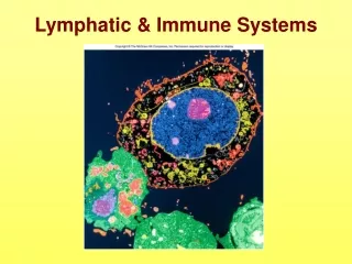
The Immune System • Recognizes specific foreign molecules • Destroys pathogens effectively • Key cells – lymphocytes • Also includes lymphoid tissue and lymphoid organs
Lymphocytes • Infectious organisms attacked by inflammatory response • macrophages, then lymphocytes • Cytotoxic T lymphocytes • Attack foreign cells directly • Binds to antigen-bearing cells • Perforates cell membrane • Signals cell to undergo apoptosis
Lymphocytes • B lymphocytes - become plasma cells - secrete antibodies, mark cells for destruction by macrophages
Lymphocyte Activation • Lymphocytes originate in bone marrow • T lymphocytes travel to the thymus gland • B lymphocytes stay in bone marrow • Able to recognize a unique antigen • Gain immunocompetence • travels through blood stream • meets and binds to a specific antigen
Lymphocyte Activation • Activating T or B cells produce • Effector lymphocytes • Short-lived, attack immediately • Memory lymphocytes • Wait until body encounters their antigen again • Basis of acquired immunity • Guard against subsequent infections
Lymphoid Tissue • Most important tissue of the immune system Two general locations: • Mucous membranes of digestive, urinary, respiratory, and reproductive tracts - Mucosa-associated lymphoid tissue (MALT) • Lymphoid organs (except thymus)
Lymphoid Organs • Primary lymphoid organs • Bone marrow • Thymus • Secondary lymphoid organs • Lymph nodes, spleen, tonsils • Aggregated lymphoid nodules • Appendix
Lymphoid Organs • Designed to gather, destroy infectious microorganisms Figure 20.10
Thymus • Immature lymphocytes develop into T lymphocytes - secretes thymic hormones - most active in childhood • Functional tissue atrophies with age - composed of cortex and medulla - medulla contains Hassall’s corpuscles (thymic corpuseles) • Differs from other lymphoid organs - functions strictly in lymphocyte maturation - arises from epithelial tissue
Thymus Figure 20.11
Lymph Nodes Functional pathway • Lymph percolates through lymph sinuses • Most antigenic challenges occur in lymph nodes • Antigens destroyed – activate B and T lymphocytes
Spleen • Largest lymphoid organ • Two main blood-cleansing functions • Removal of blood-borne antigens • Removal and destruction of old or defective blood cells • Site of hematopoiesis in the fetus
Spleen • Destruction of antigens • Site of B cell maturation into plasma cells • Phagocytosis of bacteria and worn-out RBCs, WBCs and platelets • Storage of platelets
White pulp – thick sleeves of lymphoid tissue • Red pulp - surrounds white pulp - composed of venous sinuses - splenic cords
Spleen Figure 20.12
Tonsils • Simplest lymphoid organs • Four groups of tonsils • palatine, lingual, pharyngeal, and tubal tonsils • Arranged in a ring to gather and remove pathogens • Underlying lamina propria consists of MALT
Palatine Tonsil Figure 20.13
Aggregated Lymphoid Nodules and Appendix • MALT – abundant in walls of intestines • Fight invading bacteria • Generate a wide variety of memory lymphocytes - aggregated lymphoid nodules (Peyer’s patches) - located in the distal part of the small intestine • Appendix – tubular offshoot of the cecum
Aggregated Lymphoid Nodule Figure 20.14
Disorders of the Lymphatic and Immune Systems • Chylothorax - leakage of fatty lymph into the thorax • Lymphangitis - inflammation of a lymph vessel • Mononucleosis - caused by Epstein-Barr virus - attacks B lymphocytes • Hodgkin’s disease - malignancy of lymph nodes • Non-Hodgkin’s lymphoma- uncontrolled multiplication - metastasis of undifferentiated lymphocytes
The Lymphatic and Immune Systems Throughout Life • Lymphatic vessels and lymph nodes • develop from lymphatic sacs • Thymus originates as an outgrowth of endoderm • Spleen, lymph nodes, and MALT • arise from mesodermal mesenchyme