Download
1 / 26
260 likes | 359 Views
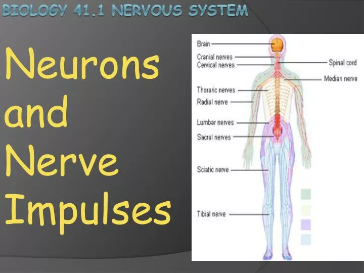
Neurons and N erve Impulses. Biology 41.1 nervous System. If your body used only chemical signals to send messages, your interaction with the environment would be very slow, t oo slow to respond with movement.

E N D
Neurons and Nerve Impulses Biology 41.1 nervous System
If your body used only chemical signals to send messages, your interaction with the environment would be very slow, too slow to respond with movement. A quicker means of communication is needed, especially if your brain has an urgent message for the muscles in your legs like “ run for your life!” I n addition to chemical signals, your nervous system uses electrical energy to send signals rapidly throughout your body. neurons
The nervous system contains a complex network of nerve cells, or neurons. Neurons are specialized cells that transmit information throughout the body. Neurons enable many important things , such as movement, perception, thought, emotion and learning. Neurons
A neuron’s unique structure enables it to conduct electrical signals called nerve impulses. Neurons communicate by transmitting nerve impulses to body tissues and organs, including muscles, glands, and other neurons. Neurons vary greatly in form, but 4 basic types of neurons are shown at right. neurons
Dendrites, which extend from the cell body of the neuron, are the antennas of the neuron. Dendrites receive information from other cells. The neuron’s cell body collects information from other dendrites, relays this information to other parts of the neuron, and maintains the general function of the neuron. dendrites
An axon is a long membrane covered extension of the cytoplasm that conducts nerve impulses. The ends of an axon are called axon terminals. When a neuron communicates with other cells, it does so at its axon terminals. Axons
Nervous tissue consists mostly of neurons and their supporting cells. Bundles of axons are called nerves. The arrangements of axons in a nerve is similar to a telephone cable with many different communication channels, each carried by a separate wire. Nerves appear as fine, white threads when viewed with the unaided eye. Nerves
Many neurons have a layer of insulation on their axon called a myelin sheath. Myelin is produced by supporting cells that surround the axon. The presence of myelin causes nerve impulses to move faster down the axon. The myelin sheath is interrupted at intervals, leaving gaps called nodes of Ranvier, where the axon membrane is exposed to the surrounding fluid. Myelin sheath
Conduction of nerve impulses is faster in myelineated axons because nerve impulses “jump” from node to node as they move down the axon. Myelin is especially beneficial in neurons that must transmit information very rapidly, such as those involved in muscle movement. Myelineated axons
All cells have an electrical charge on the inner surface of the cell membrane that is different than the electrical charge of the fluid outside the cell. The difference in electrical charge across the cell membrane, called the membrane potential, results from the movement of chargedions into and out of the cell. Neuron Function
This movement depends on the relative concentration of ions inside and outside the cell, the ability of the ions to diffuse across the cell membrane, and the electrical charges of the ions. The membrane potential is expressed as voltage, like that of a battery. Membrane potential
Ions diffuse across a neuron’s cell membrane by passing through proteins that act as ion channels. Each type of channel allows only specific ions to pass. Certain channels are voltage gated, they open or close depending on the membrane potential, apositive (+) or negative charge (-) . Even a small change in the membrane potential can affect the permeability of the cell membrane to certain charged ions. As these ions diffuse in and out of the neuron, they can affect the membrane potential. Membrane potential
When a neuron is conducting a nerve impulse, the neuron is said to be at rest. The membrane potential of a neuron at rest is called the resting potential. In a typical neuron, the resting potential is negative, about -70 mV. At the resting potential, the inside of the cell is negatively (-) charged with respect to the outside of the cell. Resting Potential
Why is the resting potential negative? Recall that sodium-potassium pumps actively (active transport=uses energy) transports sodium ions out of a cell and potassium ions into a cell. This results in a greater concentration of sodium atoms (Na+) outside the cell than inside the cell, and a greater concentration of potassium ions (K+) inside the cell than outside it. In a neuron, voltage-gated sodium channels are closed at the resting potential. Thus, very few sodium atoms can diffuse into a cell, despite the strong concentration gradient. Resting Potential
Some voltage-gated potassium channels are open at the resting potential. Potassium ions can therefore diffuse out of the cell down their concentration gradient, carrying their positive charge with them. Neurons also contain negatively charged proteins that are too large to exit the cell through channels. Resting Potential
When a neuron is conducting a nerve impulse, changes occur in the cell membrane of the neuron. A nerve impulse is also called an action potential. An action potential is a local reverse of polarity (from a negative to a positive charge) inside the neuron. An action potential moves down an axon like a flame burning down a fuse. Action Potential
A junction at which a neuron meets another cell is called a synapse. At synapses, neurons usually do not touch the cells they communicate with. Between an axon terminal and a receiving cell is a tiny gap called a synaptic cleft. At a synapse, the transmitting neuron is called a presynaptic neuron, and the receiving cell is called a postsynaptic cell. Communication between neurons
When a neuron impulse arrives at an axon terminal of a presynaptic neuron, the impulse cannot cross the synaptic cleft. Instead, the impulse triggers the release of signal molecules called neurotransmitters into the synaptic cleft. Neurotransmitter molecules are produced by neurons and are stored inside vesicles. neurotransmitters
There are many different neurotransmitters and several mechanisms of neurotransmitter action. For example, in human muscles the principal neurotransmitter is a chemical called acetylcholine. The brain utilizes several neurotransmitters such as glutamate and dopamine. neurotransmitters
A nerve impulse causes a presynaptic Neuron to release neurotransmitter molecules into the synaptic cleft. When an action potential reaches an axon terminal of the presynaptic neuron, vesicles that contain neurotransmitter molecules fuse with the cell membrane. This releases neurotransmitter molecules into the synaptic cleft by exocytosis. Release of neurotransmitter
Neurotransmitter molecules diffuse across the synaptic cleft and interact with the postsynaptic cell. Neurotransmitter molecules bind to receptor proteins on the postsynaptic cell. In some cells, ion channels open when a neurotransmitter binds to these receptor proteins. Such channels are called chemical-gated ion channels; whether these channels are open or closed depends on the binding of the chemical – in this case a neurotransmitter molecule. Release of neurotransmitter
A neurotransmitter may either excite or inhibit the activity of the postsynaptic cell it binds to. For example, when the neurotransmitter opens chemically gated ion channels, ions move across the cell membrane of the postsynaptic cell. This causes the membrane potential of the postsynaptic cell to change depending on the charge of the ions that move into or out of the cell. Release of neurotransmitter
If positively charged ions move into a postsynaptic neuron, an action potential may be produced (excitation). On the other hand, if positively charged ions flow out of the postsynaptic neuron, an action potential may be suppressed (inhibited). Release of neurotransmitter
Neurotransmitter molecules do not remain in the synaptic cleft indefinitely. Instead, most neurotransmitter molecules are cleared from the synaptic cleft very soon after they are released. Many presynaptic neurons reabsorb neurotransmitter molecules and use them again. Neurotransmitter molecules
At other synapses, neurotransmitter molecules are broken down by enzymes or other chemicals. This happens, for example, at the synapses between neurons and skeletal muscle cells. The reuptake or breakdown of the neurotransmitter molecules ensures that their effect on postsynaptic cells is not long lasting. Neurotransmitter molecules