Download
1 / 32
330 likes | 631 Views
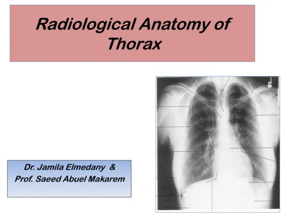
Balasubramanian Thiagarajan. Radiological anatomy of Frontal sinus. Introduction. Highly complex and variable anatomy Variations – impact on drainage Efficiency of muco-ciliary clearance – relationship to morphology of frontal sinus. Embryology. Embryology - contd.

E N D
Balasubramanian Thiagarajan Radiological anatomy of Frontal sinus drtbalu's otolaryngology online
drtbalu's otolaryngology online Introduction • Highly complex and variable anatomy • Variations – impact on drainage • Efficiency of muco-ciliary clearance – relationship to morphology of frontal sinus
drtbalu's otolaryngology online Embryology
drtbalu's otolaryngology online Embryology - contd • Continuation of embryonic infundibulum, frontal recess superiorly • Upward migration of anterior ethmoid air cells • Penetration via inferior aspect of frontal bone between the two tables • Pneumatization 2-9 years • Complete – 9 years
drtbalu's otolaryngology online Frontal sinus drainage • Expands into the diploic space of frontal bone from frontal sinus ostium • Each sinus grows independently of the other • Growth is dependent on ventilation, drainage, growth of surrounding sinuses and skull base • Drainage is hence highly variable
drtbalu's otolaryngology online Frontal sinus drainage - contd • Inferomedially into funnel shaped area (frontal sinus ostium) • Anterior wall of drainage channel is bounded by nasal beak • Ostium is oriented perpendicular to the posterior wall of the sinus at the skull base level
drtbalu's otolaryngology online Frontal beak
drtbalu's otolaryngology online Frontal sinus drainage pathway compartments • Drainage pathway is divided into superior and inferior compartments • Superior compartment is formed by union of adjacent air cells at the antero inferior portion of ethmoid bone • Size and shape of this component varies with the varying anatomy for fronto ethmoidal air cells • Superior compartment communicates with the inferior compartment
drtbalu's otolaryngology online Inferior component • This passage is really narrow • This compartment is formed by ethmoidal infundibulum / middle meatus • This is dependent on the attachment of uncinate process
drtbalu's otolaryngology online Inferior compartment Inferior portion - infundibulum Inferior portion - infundibulum
drtbalu's otolaryngology online Attachment to lamina papyracea – inferior component formed by middle meatus
drtbalu's otolaryngology online Inferior compartment formed by infundibulum
drtbalu's otolaryngology online Frontal beak Frontal beak forms the floor of frontal sinus
drtbalu's otolaryngology online Frontal recess • Antero superior portion of ethmoidal air cell system • This is where the frontal bone pneumatization begins • Lateral wall is formed by lamina papyracea • Medial wall is formed by vertical attachment of middle turbinate • Posterior wall is variable ? Bulla if it manages to reach up to skull base
drtbalu's otolaryngology online Frontal recess
drtbalu's otolaryngology online Agger nasi
drtbalu's otolaryngology online Large bulla • Can obstruct frontal sinus outflow • This area should be critically studied during imaging • Causes obstruction from behind
drtbalu's otolaryngology online Uncinate vs frontal sinus drainage • Uncinate – skull base attachment causes frontal sinus to drain into superior portion of ethmoidal infundibulum • Uncinate – attached to middle turbinate causes frontal sinus to drain into ethmoidal infundibulum • Uncinate – attached to lamina papyracea causes frontal to drain into superior aspect of middle meatus. Ethmoidal infundibulum ends in terminal recess
drtbalu's otolaryngology online Ethmoidal infundibulum • 3 – d space • Lateral – LP • Anteromedial – Uncinate • Posterior - Bulla
drtbalu's otolaryngology online Frontal recess block Large agger cell causing narrowing of frontal recess
drtbalu's otolaryngology online uncinate process attached to skull base. The frontal recess is seen between the agger nasi and uncinate process
drtbalu's otolaryngology online Scan showing uncinate process being attached to lamina papyracea. This causes terminal recess to form. Frontal sinus drains directly into middle meatus
drtbalu's otolaryngology online scan shows the uncinate process being attached to the middle turbinate. Note the presence of infundibulum between the bulla and the uncinate process. Frontal sinus is seen opening into the infundibulum
drtbalu's otolaryngology online Bent's classification of frontal cell variants • Type I: Single frontal recess cell above the agger • Type II: Tier of air cells above agger projecting into the frontal recess • Type III: Single massive air cell above agger expanding in superior direction • Type IV: Single isolated cell within frontal sinus. Difficult to visualize due to thin walls
drtbalu's otolaryngology online Type I & II frontal cell
drtbalu's otolaryngology online Type III
drtbalu's otolaryngology online Type IV
drtbalu's otolaryngology online Supraorbital air cells • Pneumatization of orbital plate of frontal bone • Posterior to frontal recess & lateral to frontal sinus • Sometimes can reach high up mimicking frontal sinus
drtbalu's otolaryngology online Thank you