Download
1 / 42
420 likes | 455 Views
Self explanatory slides on the branches of the Abdominal aorta radiologically

E N D
Radiological anatomy of the abdominal aorta and its major branches. by Dr Taiwo Tosin Ojeremi Department of Radiology, Ahmadu Bello University Teaching Hospital Shika- Zaria Nigeria
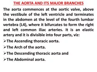
Introduction • Continuation of thoracic aorta at diaphragmatic hiatus(12th thoracic vertebra) • It is 12cm long,2-3cm wide • It lies anterior to T12/l1 to l4 vertebrae • Accompanied by the thoracic duct, azygous and hemi-azygous vein, the sympathetic trunk and splanchnic nerve.
Branches of the abdominal aorta seen on angiogram as follows: • Three unpaired anterior branches: Coeliac trunk at T12/L1 Superior mesenteric artery at L1 Inferior mesenteric artery at L3 • Three lateral paired visceral arteries: Adrenal arteries at L1 Renal arteries at L1/L2 Gonadal arteries at L3
Cont’d • Five lateral paired parietal branches: Inferior phrenic arteries at T12 Four pairs of lumbar arteries • Terminal arteries: The common iliac arteries Median sacral artery.
Radiological anatomy of abdominal aorta and its major branches
Doppler ultrasound of the Aorta • Sharp increase antegrade flow velocity followed • Rapid decrease that bottoms out in early diastole with a brief period of reversed flow. • Low velocity antegrade flow then resumes and continue for the rremainder of diastole.
CT at supra renal level demonstrating abd. Aorta and it branches
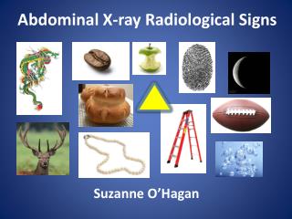
Plain films • Uses ionizing radiation though cheap and readily available • The abd. Aorta and its major branches are not demonstrable on plain radiograph except if calcified • if calcified, It is then seen as linear calcification vertically in the midline and to the left. .