Download
1 / 4
40 likes | 42 Views
Virchowu2019s node, left supraclavicular lymph node contains metastases of many thoracic and abdominal visceral malignancies such as lung, breast, esophageal, gastric, pancreatic cancers. Metastasis to non-regional lymph nodes especially cervical lymph nodes is extremely rare presentation as in this case.

E N D
Journal Home Page www.bbbulletin.org BRITISH BIOMEDICAL BULLETIN Case Report Virchow’s Node: A Look Beyond Gut Carcinoma C. Bharath*1, Komala H M2 1Prof & Head, Dept. of Pathology,Vijayanagar Institute of Medical Sciences, BELLARY. Karnataka. India. 2Post Graduate, Vijayanagar Institute of Medical Sciences, BELLARY. Karnataka. India. A R T I C L E I N F O Received 07 Dec. 2013 Received in revised form 12 Dec. 2013 Accepted 20 Dec. 2013 Keywords: Urothelial carcinoma; Supraclavicular lymphnode; Metastasis; Rare entity, Differential diagmosis. Corresponding author:Prof & Head, Dept. of Pathology,Vijayanagar Institute of Medical Sciences, BELLARY. Karnataka, India E-mail address: bhar5anu@yahoo.co.in A B S T R A C T Virchow’s node, left supraclavicular lymph node contains metastases of many thoracic and abdominal visceral malignancies such as lung, breast, esophageal, gastric, pancreatic cancers. Metastasis to non-regional lymph nodes especially cervical lymph nodes is extremely rare presentation as in this case. A middle aged male on examination found left supraclavicular lymphnodes enlaragement and subjected for FNAC. On microscopy the features were suggested of metastatic urothelial deposits histopathology. As this is a rare metastatic presentation at this lymph node, one shood keep in mind this as a differential diagnosis for supraclavicular lymph node enlargement. & confirmed by ©2015 British Biomedical Bulletin. All rights reserved
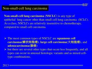
Bharath et al____________________________________________________ ISSN-2347-5447 Introduction Virchow’s node, left supraclavicular lymph node contains metastases of many thoracic and abdominal malignancies such esophageal, gastric, pancreatic, gynecologic, and prostate cancers2. Urothelial carcinoma accounts for 90% of cases of bladder cancer with metastases usually limited to the regional pelvic nodes4.Metastasis to non- regional lymph nodes especially cervical lymph nodes is presentation1.Only few reports have been published so far and with poor prognosis1. Though distant lymph node involvement is rare but cannot be entirely overlooked. Case report revealed primary tumor to be Urothelial carcinoma. (Fig.3) Supraclavicular lymph node metastases are rare in this case and indicate widespread disease with poor prognosis. Discussion visceral breast, as lung, Bladder cancer is the most common malignant disease of the urinary tract4.It is commonly a disease of older age and is more prevalent among men than women4.It is the 2nd most prevalent cancer for men and 10thmost prevalent cancer for women3.It has variable metastatic potential and almost any organ can be involved.Data on its metastatic pattern are limited.4 The pattern of recurrence and metastases are not dependent on the features of the primary tumor5. Common sites of metastatic spread of bladder carcinoma are regional lymph nodes (90%), liver (47%),lung (45%), bone (32%), peritoneum (19%), pleura(16%), kidney (14%), adrenal gland (14%), and the intestine (13%)2..The most common lymph nodes involved are external, internal iliac and obturator (20%-45%) lymphatic drainage of the bladder and the common iliac sites as the secondary drainage1. The possible route of spread to head and neck region is by haematogenous through vertebral veins and by lymphatics1. Presence of Virchow’s node invasivebladder tumour is considered as incurable metastatic pathological retrograde tumour cell deposition against the normal drainage of the node (towards the thoracic duct) imply extensive tumour occupation of the retro peritoneum. Study done by Hessan et al. among 207 patients with metastasis to the head and neck area lymph nodes showed only 3 cases having metastasis Urothelial in origin5.Ferlito et al reported a series of genitourinary tumors and found this group to be the third most extremely rare A 40 year old male presented with history of fever, intermittent hematuria and burning micturition Incidentally left supraclavicular lymph node was found to be enlarged. Patient was subjected for FNAC. Results since one week. as the primary FNAC showedcellular smears consisting of atypical epithelial cells in papillary fragments,monolayered sheets and loose clusters with both squamous and glandular differentiation. These cells showed stratification of the nuclei within the fragments. Cells with eccentrically placed nucleus, spindle cells, racquet like cells, pyramidal cells, atypical stripped nuclei were also seen. It was diagnosed as metastasis of Urothelial carcinoma (Fig.1 and Fig.2). Patient was subjected to further relevant investigations. CT scan showed well defined enhancing mass lesion in the bladder measuring 4.5×4.8 cm arising from anterior wall with intraluminal Hypodenselesions in both lobes of liver and right iliac fossaseen suggestive of metastases. Biopsy of the bladder mass was done which with muscle disease as the extension. BBB[3][2][2015] 199-202
Bharath et al____________________________________________________ ISSN-2347-5447 2. frequent tumor site to metastasize to the supraclavicular fossa3. Conclusion L. N. Seneviratne, J. M. N. R. K. Jayasundare and N. D. Perera, Virchow’s node – an unheard site of metastatic bladder cancer: Sri Lanka Journal of Urology: 2009: 10:28-30 Ore Ogunyemi · A. Rojas · K. Hematpour ,D. Rogers · C. Head · C. Bennett, Metastasis of genitourinary tumors to the head and neck Otorhinolaryngol (2010) 267:273–279 Atul B. Shinagare1,Nikhil H. Ramaiya, Jyothi P. Jagannathan,Fiona M. Fennessy, Mary-Ellen Taplin, Annick D. Van den Abbeele:Metastatic Pattern of Bladder Cancer: Correlation With the Characteristics of the Primary Tumor: AJR 2011; 196:117– 122 VenkatPavanKancharlaa GulmibArefAghelia,MichaelDegenbArashG oharic Ming Jiangd J.C. Wanga: Transitional Cell Carcinoma of the Bladder Manifestating as Malignant Lymphoma with Generalized Lymphadenopathy: 2010:3:125–130. 3. This case is a rare presentation of Urothelial carcinoma metastases to Virchow’s node. Identification of nodal involvement is important because the presence of nodal metastasis advances the disease to stage IV4. Picking up nodal metastases may influence therapeutic decisionsand FNAC can be used as first line investigation in diagnosing such metastases with certainty. References 1. Mutahir A. Tunio, Mushabbab A lAsiri, Yasser Bayoumi, MohsinFareed, Shoaib Ahmad, Cervical lymph node metastasis from transitional cell carcinoma of urinary bladder: Case report and review of literature: Journal of Solid Tumors: June 2012:Vol. 2: Issue 3:59-62 region: Eur Arch 4. 5. Frederick A. Case Rep Oncol Figure 1. FNAC showing atypical epithelial cells arranged in papillary fragments(H&E 10x) BBB[3][2][2015] 199-202
Bharath et al____________________________________________________ ISSN-2347-5447 Figure 2. Cluster ofpleomorphic cells showing nuclear overlapping with coarse chromatin (H&E 45x) Figure 3. Atypical tumor cells in varied pattern diagnosed as urothelial carcinoma (H&E 40x) BBB[3][2][2015] 199-202