Download
1 / 42
500 likes | 1.25k Views
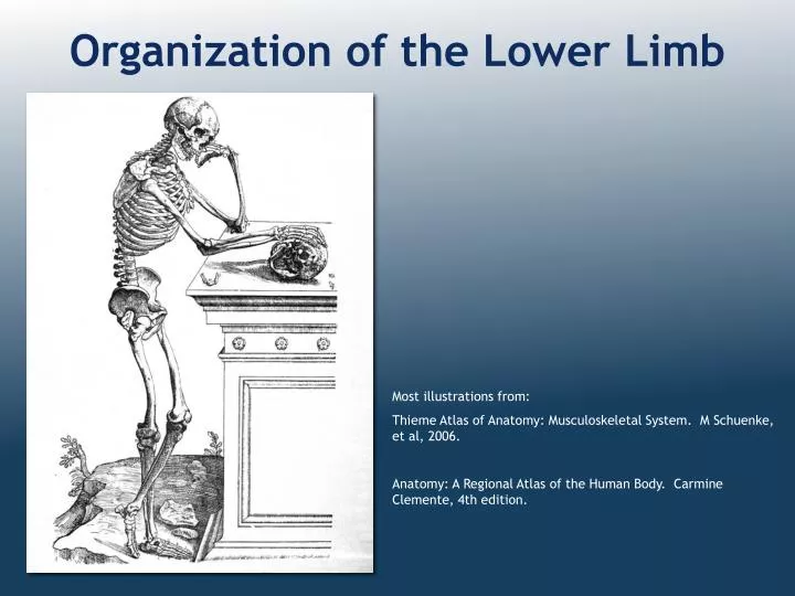
Organization of the Lower Limb. Most illustrations from: Thieme Atlas of Anatomy: Musculoskeletal System. M Schuenke, et al, 2006. Anatomy: A Regional Atlas of the Human Body. Carmine Clemente, 4th edition. Limb Development.

E N D
Organization of the Lower Limb Most illustrations from: Thieme Atlas of Anatomy: Musculoskeletal System. M Schuenke, et al, 2006. Anatomy: A Regional Atlas of the Human Body. Carmine Clemente, 4th edition.
Limb Development • Lower limb develops in an anterolateral position at the level of the L2 to S3 trunk segments • Great toe positioned in cephalic direction with the soles of the feet in an ateriomedial direction • Knee positioned in a posteriolateral position
Limb Rotation Lower Limb Rotation • Medial rotation • Adduction • Extension
Segments of the Lower Limb • Limb Girdle • pelvic girdle (gluteal) • Free limb • thigh • leg • foot
Articulation with Axial Skeleton Sacroiliac jt. Sternoclavicular jt,
Sacroiliac Joint Auricular articular surfaces
Sacroiliac Joint Anterior sacroiliac ligaments Sacroiliac joint Posterior sacroiliac ligaments
Os Coxae • Hemipelvis is composed of the ilium, ischium and pubis with articulations centered in acetabulum – triradiate articulation • Fusion of the 3 bones is complete between 20 and 25 years of age
Joints of the Free Limb • Hip • Polyaxial ball and socket joint: • Felxion/extension • Adduction/abduction • Medial/lateral rotation Iliofemoral lig. Pubofemoral lig. Ischiofemoral lig. Ligament of head of femur
Joints of the Free Limb Knee Modified hinge joint Tibiofemoral artic. Patelofemoral artic. Femur Tibia Patella
Joints of the Free Limb Ankle (talocrural) joint Hinge joint 1 axis of movement: Dorsiflexion/plantarflexion
Joints of the Free Limb Foot Interphalangeal joints: Distal Proximal metatarsophalangeal joints Inversion/eversion occurs at subtalar (talocalcaneal) joint
Superficial Veins Great saphenous v. Small saphenous v.
Arterial Blood Supply External Iliac a Femoral a Popliteal a Anterior Posterior tibial a tibial aa dorsalis med/lat pedis plantar a Internal Iliac a Superior gluteal a Inferior gluteal a Obturator a
Deep Femoral Artery Medial & lateral femoral circumflex arteries
Fascia of the Lower Limb Gluteal Fascia Fascia Lata Crural Fascia
Fascial Compartments of the Thigh Fascial septa divide limb into compartments anterior compartment medial compartment posterior compartment Anterior compartment Medial (adductor) compartment Posterior compartment Fascia Lata
Fascial Compartments of the Leg anterior compartment Anterior crural compartment Lateral crural compartment Posterior crural compartment lateral compartment posterior compartment anterior lateral Superficial & deep posterior crural compartments Crural Fascia posterior
Lumbosacral Plexus L1 - S3
Gluteal innervation: • Superior gluteal n (L4,5, S1): • gluteus medius m. • gluteus minimus m. • tensor fascia lata • Inferior gluteal n (L5, S1,2): • gluteus maximus m. • N to obturator internus(L5, S1, 2): • oburator internus • superior gemellus • Nerve to quadratus femoris(L4,5, S1): • inferior gemellus • quadratus femorus
Compartmental Innervation Of the Free Limb Anterior compartment Medial compartment Posterior compartment Lateral compartment Thigh Leg Foot
Anterior (extensor) compartment of the thigh Innervation: femoral nerve anterior anterior
Medial (adductor) compartment of the thigh Innervation: obturator nerve* medial *Adductor Magnus innervated by both obturator n and tibial division of sciatic n medial
Posterior compartment of the thigh Hamstring mm: arise from ischial tuberosity & are innervated by tibial division of sciatic n. • Innervation: • Tibial division of sciatic nerve • Except short head of biceps femoris: common fibular division of sciatic nerve posterior
Posterior compartment of the leg Innervation: tibial n. posterior Superficial posterior crural compartment Deep posterior crural compartment
Plantar surface of the foot Innervation: Medial & lateral plantar nerves
Innervation of intrinsic muscles of the foot Medial plantar n •flexor digitorum brevis •flexor hallucis brevis •abductor hallucis m •1st lumbrical Lateral plantar n •abducator digiti minimi •quadratus plantae •2nd, 3rd & 4th lumbricals •adductor hallucis •Flexor digiti minimi •dorsal & plantar interossei
Common fibular nerve Nerve supply to the lateral and anterior crural compartments anterior lateral anterior lateral
Lateral crural compartment: Superficial fibular nerve Fibularis longus Fibularis brevis lateral
Anterior crural compartment: • Deep fibular nerve • Tibialis anterior m. • Extensor hallucis longus m. • Extensor digitorum longus m. • Fibularis tertius m. anterior
Dorsum of the foot: • deep fibular nerve • Extensor hallucis brevis • Extensor digitorum brevis
Compartmental Innervation of Free Limb Anterior compartment Medial compartment Posterior compartment Lateral compartment Tibial division of sciatic n. * Thigh Femoral n. Obturator n.* *adductor magnus - obturator n and tibial n *short head of biceps femoris - common fib. division of sciatic n. Leg Superficial fibular n. Deep fibular n. Tibial nerve Foot Deep fibular n. Tibial n. (medial and lateral plantar n.)
Dermatomes • Key dermatomes* • thumb - C6 • 5th digit of hand - C8 • nipples - T4 • umbilicus - T10 • hip crease - L1 • great toe - L4 • 5th toe - S1 • *area of skin supplied by a dorsal nerve root
Key dermatomes* of the Lower Limb • umbilicus - T10 • hip crease - L1 • great toe - L4 • 5th toe - S1 • *area of skin supplied by a dorsal nerve root
References Clemente, C. 1997 Anatomy: A Regional Atlas of the Human Body, 4th edition. Lippincogtt Williams & Wilkins. Rinabesm C,H, 1972. Cunningham’s Textbook of Anatomy, 11th edition. Oxford University Press. Rosse, C & Gaddum-Rosse, P. 1997. Hollinshead’s Textbook of Anatomy 5th edition. Lippincott-Raven Schuenke, M et al, 2006 Thieme Atlas of Anatomy: Musculoskeletal System. Thieme. Slaby, F., et al. 1994. Gross Anatomy in the Practice of Medicine. Lea & Febiger.