Download
1 / 27
270 likes | 395 Views
A1.2CM1 Cells, Molecules & Metabolism Lecture 25 (2002) Professor M Schweizer Refer to chapter 18, Stryer, 5e. Cellular respiration. Karp 3e, Figure 5.5. Figure 5.2c. Confocal microscopy reveals mitochondrion as sausage. was. is now. Electron Flow produces heat, light, movement, etc.

E N D
A1.2CM1 Cells, Molecules & Metabolism Lecture 25 (2002) Professor M Schweizer Refer to chapter 18, Stryer, 5e Cellular respiration
Confocal microscopy reveals mitochondrion as sausage was is now
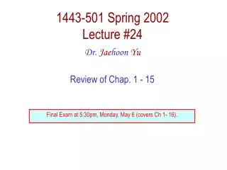
Electron Flow produces heat, light, movement, etc Bio-battery Bio-wire is the respiratory assembly; electron flow produces ATP; drawn by Dr B J Catley)
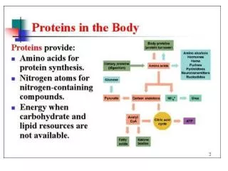
Electron Transfer An electron transfer reaction: Aox + Bred Ared + Box Aox is the oxidized form of A (the oxidant) Bred is the reduced form of B (the reductant). One may consider as two half-cell reactions: Aox + n e- Ared e.g., Fe+++ + e- Fe++ Box + n e- Bred
Aox + n e- Ared Box + n e- Bred For each half reaction: E = E°'– RT/nF (ln [reduced]/[oxidized]) e.g., for the first half reaction: E = E°' – RT/nF(ln[Ared]/[Aox]) E = voltage, R = gas const., F = Faraday (23.06 kcalmol-1 V-1), n = # e-. When [Ared] = [Aox], E = E°'. E°' is the mid-point potential, or standard redox potential, the potential at which [oxidant] = [reductant] for the half reaction. Standard redox potential for the oxydation-reduction reaction involving hydrogen (2H+ + 2 e H2) E0 = 0.00V (H+ = 1.0 M (pH=0)); E0’ = -0.42 V (H+ = 10-7 M (pH = 7))
ElectronTransfer Consider transfer of 2 electrons from NADH to oxygen: a. ½ O2 + 2H+ + 2e- H2O E°' = +0.815 V b. NAD+ + 2H+ + 2e- NADH + H+ E°' = -0.315 V Subtracting reaction b from a: c. ½ O2 + NADH + H+ H2O + NAD+ DE°'= +1.13 V DG = - nFDEo' = – 2(96494)(1.13) = -52.6 kcal mol-1 (-218 kJ mol-1)
ElectronTransfer Consider transfer of 2 electrons from H2 to oxygen: a. ½ O2 + 2H+ + 2e- H2O E°' = +0.815 V b. 2H+ + 2e- H2 E°' = -0.42 V Subtracting reaction b from a: c. ½ O2 + H2 H2O DE°'= +1.23 V DG = - nFDEo' = – 2(96494)(1.13) = -57 kcal mol-1 (-239.4 kJ mol-1)
Electron Transfer For an electron transfer: DE°' =E°'(oxidant) – E°'(reductant) = E°'(acceptor) – E°'(donor) DGo' = – nFDE°' (E°' is the mid-point potential) An electron transfer reaction is spontaneous (negative DG) if E°' of the donor is more negative than E°' of the acceptor, i.e., when there is a positive DE°'.
ElectronCarriers NAD+/NADH and FAD/FADH2 were introduced earlier. FMN (Flavin MonoNucleotide) is a prosthetic group of some flavoproteins. It is similar in structure to FAD (Flavin Adenine Dinucleotide), but lacking the adenine nucleotide. When free in solution, FMN (like FAD) can accept 2 e- + 2 H+ to form FMNH2.
Role of FMN Role of FMN mediating between 2e- & 1e- carriers: For example, when NADH donates electrons to the respiratory chain, the initial electron transfers are: NADH + H+ + FMNNAD+ + FMNH2 FMNH2 + Fe+++FMNH· + Fe++ + H+ After Fe++ is reoxidized, by passing the electron to the next carrier in the pathway: FMNH· + Fe+++FMN + Fe++ + H+
Enzyme-bound FMN can accept 1 e- to form the half-reduced semiquinone radical. The semiquinone can accept a 2nd e- to yield FMNH2. Since it can accept/donate 1 or 2 e-, FMN has an important role in mediating e- transfer between carriers that transfer 2 e- (e.g., NADH) and those that transfer 1 e- (e.g., Fe+++).
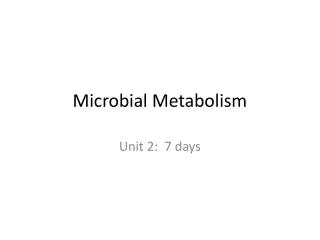
Iron-sulfur Centers Iron-sulfur centers transfer only one electron, even if they contain two or more iron atoms, because of the close proximity of the iron atoms. E.g., a 4-Fe center might cycle between redox states: Fe+++3,Fe++1(oxidized)+1 e-Fe+++2, Fe++2(reduced)
(A) A single iron ion bound by 4 cys residues (a) [2Fe-2S]; (b) [4Fe-4S] Karp 3e, Figure 5.12
Cytochromes Cytochromesabsorb light at characteristic wavelengths. Absorbance changes upon oxidation/reduction of the heme iron provide a basis for monitoring redox state. Some cytochromes are part of large integral membrane complexes, each consisting of several polypeptides and multiple electron carriers. Cytochrome c is a small, water-soluble protein with a single heme group. Cytochrome c reversibly binds to integral membrane electron transfer complexes from which it receives, or to which it donates, an electron.
The porphyrin ring is planar. Heme Fe is usually bonded to 2 axial ligands, above & below the heme plane (X,Y) in addition to the 4 N of porphyrin. Axial ligands may be S or N of amino acid side-chains. Axial ligands in cyt c are Met S (yellow) and His N (blue). A heme that binds O2 may have an open (empty) axial ligand position.
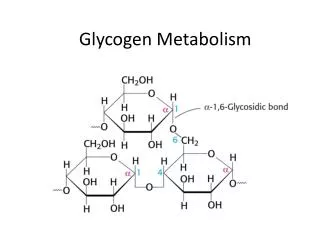
Heme is a prosthetic group of cytochromes. Heme contains an iron atom in a porphyrin ring system. The heme Fe is bonded to 4 N of the porphyrin ring.
Hemes in the 3 classes of cytochrome (a,b,c) differ in substituents on the porphyrin ring.Only heme c is covalently linked to the protein via thioether bonds to Cys residues.
Aspects of cytochrome c • structure to be explored by • Chime: • The heme sits in a crevice, with the heme plane perpendicular to the protein surface. • View the axial ligands to the heme. • On the surface of cytochrome c, positively charged lysineresidues (shown in magenta) surround the heme crevice. These are postulated to form salt bridges to anionic residues on membrane complexes to which cytochrome c binds during electron transfer.
Coenzyme Q (CoQ, Q or ubiquinone) is lipid-soluble. It dissolves in the hydrocarbon core of a membrane. Most often n = 10. The isoprene tail of Q10 is longer than the width of a lipid bilayer. CoQ has a quinone ring, which can be reduced to the quinol. Free CoQ can undergo a 2 e- oxidation/reduction: Q + 2 e- + 2 H+ QH2.
When bound to special sites in respiratory complexes, CoQ can accept 1e-to form a semiquinone radical (Q•-). CoQ functions as a lipid-soluble e- carrier, and in trans-membraneH+ transport coupled to e- transfer (Q Cycle).