Download
1 / 51
570 likes | 981 Views
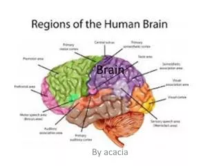
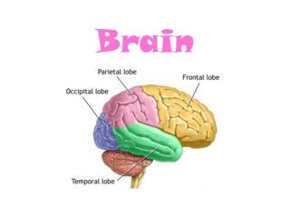
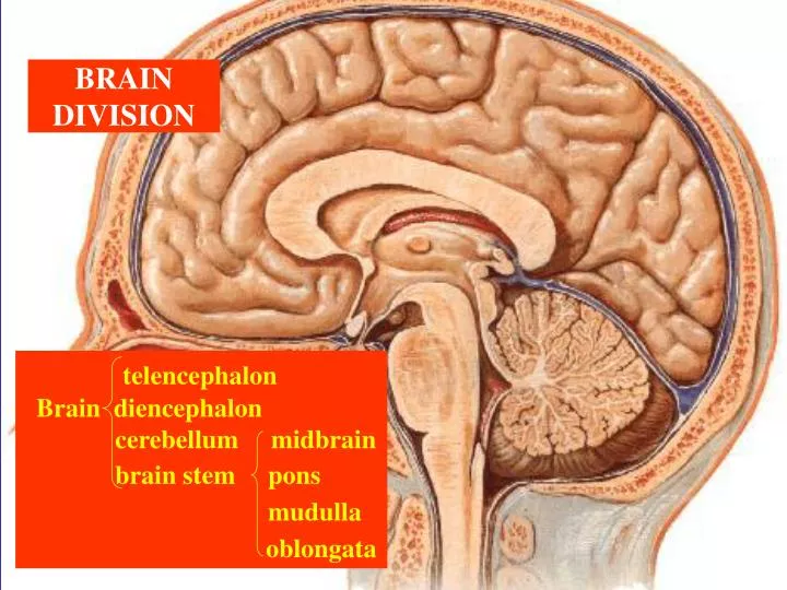
BRAIN DIVISION. telencephalon Brain diencephalon cerebellum midbrain brain stem pons mudulla oblongata. I.Brain Stem.

E N D
BRAIN DIVISION telencephalon Brain diencephalon cerebellum midbrain brain stem pons mudulla oblongata
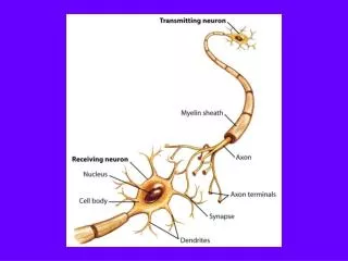
I.Brain Stem 1.Location:Being in the skull , it superiorly connects diencephalon , inferiorly conjoins spinal cord and ventrally lies on the clivus of occiptal bone and posteriorly attaches cerebellum. 2.External Borders (1) ventral surface a. bulbopotine sulcus b. sup border of basilar part in pons (2) dorsal surface a. striae medullares b. lower border of inf colliculi
3. Parts and Ventral Appearances Cerebral peduncles Interpeduncular fossa Midbrain Basilar part of pons Basilar sulcus Pons Pyramids Decussation of pyramid Inferior olives Medulla oblongata
4. Parts & Dorsal Appearances(A) Superior colliculi Brachiums of superior colliculi Inferior colliculi Brachiums of inferior colliculi midbrain Paired Structures
Rhomboid Fossa (4th ventricle floor) Parts & Dorsal Appearances(B) median sulcus 1 sulcus limitens redullary stria 1 medial eminences facial colliculi vagal-triangles hypoglossal triangles vestibular areas sup, mid & inf cerebellar peducles Pons & Medulla Oblongata Paired structures pai
3) Names of Cranial Nerve(CN) I olfactory n II optic n Ⅲ oculomotor n Ⅳ trochlear n Ⅴ trigeminal n Ⅵ abducent n (dorsal origin) Ⅶ facial n Ⅷ vestibulocochlear n Ⅸ glossopharyngeal n Ⅹ vagus n Ⅺ accessory n Ⅻ hypoglossal n
Connections with Parts Ⅲ oculomotor n Ⅳ trochlear n Midbrain Ⅴ trigeminal n Ⅵ abducent n Pons Ⅶ facial n Ⅷ vestibulo- cochlear n Ⅸ glosso- pharyngeal n Medulla oblongata Ⅹ vagus n Ⅺ accessory n Ⅻ hypoglossal n
3.Internal Structure Inclusion:gray matter,white matter & reticular formation Characteristics of Gray Matter :
Think about the followimg questions: 1.What nuclei exist in midbrain,pons and medulla oblongta, respectively? 2.Which are cranial nuclei or non-cranial nuclei? 3.How many kinds of cranial nuclei in brain stem? somatic motor nuclei visceral motor nuclei visceral afferent nuclei somatic afferent nuclei
Localization of Somatic Motor Nuclei Division of Brain Stem Somatic Motor NucleiLying Immediately Lateral to Median Sulcus nucleus of oculomotor n midbrain nucleus of trochlear n pons nucleus of abducent n medulla oblongata nucleus of hypoglossal n
Nucleus of oculomotor n Top-bottom arrangement Nucleus of trochlear n Nucleus of abducent n Nucleus of hypoglossal n Dorsal surface of brain stem
Localization of General Visceral Motor Nuclei Division of Brain Stem GeneralVisceral Motor Nuclei of CN Immediately Medial to Sulcus Limitans midbrain accessory nucleus of oculomotor n pons superior salivary nucleus medulla oblongata inferior salivary nucleus dorsal nucleus of vagus n
Accessory nucleus of oculomotor n Superior salivary n Inferior salivary n Dorsal nucleus of vagus n
Localization of Special Visceral Motor Nuclei Division of Brain Stem Special Visceral Motor Nuclei of CN Ventrolateral to VMN & Medial to Sulcus Limitans midbrain motor nucleus of trigeminal n pons nucleus of facial n medulla oblongata ambiguous nucleus accessory nucleus
Motor nucleus of trigeminal n Nucleus of facial n Ambiguous nucleus Accessory nucleus
Localization ofGeneral & Special Visceral Sensory Nucleus Division of Brain Stem General & Special Visceral Sensory Nucleus of CN Immediatedly Lateral to Sulcus Limitans midbrain none none pons medulla oblongata nucleus of solitary tract
Nucleus of solitary tract
Localization ofGeneral & Special Somatic Sensory Nuclei General & Special Somatic Sensory Nuclei Lateral & Dorsalolateral to GSVSN Division of Brain Stem mesencephalic nucleus of trigeminal n midbrain cochlear nuclei pontine nucleus of trigeminal n pons vestibular nuclei medulla oblongata spinal nucleus of trigeminal n
Mesencephalic nucleus of trigeminal n pontine nucleus of trigeminal n Cochlear & vestibular nuclei Spinal nucleus of trigeminal n
Localization of Non-Crainal Nuclei Division of Brain Stem Nuclei of Non-Cranial Nerves nucleus of sup. colliculus nucleus of inf. colliculus midbrain black substance red nucleus pons pontine nucleus gracile nucleus medulla oblongata cuneate nucleus
Test Yourself 1.Discern which one is a cranial nucleus, ambiguous or gracile nucleus? 2. Distinguish the category of the following nuclei: black substance red nucleus cochlear nuclei accessory nucleus dorsal nucleus of vagus n vestibular nuclei nucleus of hypoglossal n
3.How many nuclei are there in medullar oblongata ? 4.Which nucleus is in pons? ( memorize nuclei according to their positions) Black substance Red nucleus Cochlear nuclei Vestibular nuclei Accessory nucleus Dorsal nucleus of vagus N. Nucleus of hypoglossal N.
5.Which one is somatic nucleus? (to memorize four kinds of cranial N respectively according to their functions) Nucleus of oculomotor N Nucleus of facial N Accessory nucleus of oculomotor N Nucleus of solitary tract Spinal nucleus of trigeminal N.
6.Four pairs of parasympathetic nuclei exsit in brain stem: accessory nucleus of oculomotor n superior salivary nucleus inferior salivary nucleus dorsal nucleus of vagus n
7.Relationships between nuclei & cranial nerves1) one nucleus connects 1 nerve 2) one nucleus connects 2 or more nerves nucleus of solitary tract Ⅶ、 Ⅸ、 Ⅹ ambiguous nuleus Ⅸ、 Ⅹ、 Ⅺ
3) Two or more nuclei connect one nerve: a. oculomotor nu, accessory nu of oculomotor n---Ⅲ b. sensory & motor nus of trigeminal n---------------Ⅴ c. facial nu, sup. salivary nu, solitary tract nu------Ⅶ d. ambiguous , inf.salivary,solitary tract nus------Ⅸ e. ambiguous nu, dorsal nu of vagus n,solitary tract nu, spinal nu of trigeminal n------------- - Ⅹ
Cranial N and Relative Nuclei III IV V VIII VI VII XII IX X XI
White Matter of Brain Stem Ascending tracts: 1.medial lemniscus 2.spinal lemniscus 3.trigeminal lemniscus 4.lateral lemniscus Descending tracts: 1.corticospinal tract 2.corticonucleus tract 3.extrapyramid tract
Overview of Microstructures of White Matter 1. White matter consists of numerous nerve fibers. 2. The certain bundle fibers with the same origin, course,end & functions form a tract. 3. The fiber tracts of brain include both ascending & descending ones(ascending –blue;descending –red). 4. Focus on thenames (according to origin & end ) formations ( Which nucleus receives or sends the bundled fibers ? ), locations ( Where do the fibers pass via the brain stem ? ) and functions ( What information do the fibers transmit ? ) of the main tracts. Descending Tract
1.Medial Lemniscus medial lemniscus ↑ decussation of med lemniscus ↑ gracile nucleus cuneate nucleus ↑ fasciculus gracilis fasciculus cuneatus
2.Spinal Lemniscus anterior spinal lemniscus lateral spinal lemniscus
3.Trigeminal Lemniscus trigeminal lemniscus ↑ decussation of trigeminal lemniscus ↑ pontine nucleus & spinal nucleusof trigeminal n
4.Lateral Lemniscus lateral lemniscus ↑ ↑uncrossed ↑crossed trapezoid body ↑ cochlear nuclei
Descending Tract 1. Pyramidal Tract 1) corticonuclear tract 2) corticospinal tract 2. Extrapyramidal System
1) Corticonuclear Tract a.inf.part of precentral gyrus ↓ motor nuclei in brain stem b.The tract dominates the most nuclei on both sides of brain stem. c.Lower-half parts of facial nu & hypoglossal nu only receive the control of the tract from opposite side. d.The tract controls voluntarily muscular movements of head, face and neck .
2) Corticospinal Tract superior & middle part of precentral gyrus ↓ ant horn cells of spinal cord a.Most fibers decussate at decussation of pyramid. b.Fewer fibers decussate at ant white commissure. c.The tract controls movements of voluntary muscles in trunk and limbs.
Extrapyramidal System All the descending tracts except pyramidal tracts dominating the vuluntarily muscular movements, are subordinated to the system. The functions of the system are to modulate the voluntarily muscular movements. The conbination of the two system enables the body to carry out the appropriate, precise , effective, harmonious & temporospatial movements so as to reliably respond to the stimuli from external or internal enviroments.
Reticular Formation of Brain Stem • Raphe nuclei • locates in • median line • of brain stem. • Functions: • ascending : • helping sleep • 2) descending: • alleviating pain
Ascending Reticular Activating System--ARAS Location: dorsolateral area of pons and medullar oblongata Functions: (1)~(3)
(1)Receives the informations transmitted by the numerous lateral branches given off by the responding specific sensation conduction tracts. (2)converts these specific messages into non-specific ones ,and then sends them to most areas of cortex of cerebrum via its projection fibers. (3) thus makes the cortex be conscious or vigilant states so as to be able to recognize and distinguish the various afferent messages. Fiber projection targets of ARAS Fibers to cortex Lateral branches pons Sensory pathways bolb
a.Regulating somatic movements: strengthen or weaken muscular tensions . b.Performing the behavioral arousal function. c.Controlling the activities: vomiting center;regulating visceral reflexes(DS&RS); centers for vital movements , including cardiovascular motor center & respiratory center----controlling heartbeat, BP& R. d.5-TH ascending projection to cerebral cortex inhibits activities of the cortex and induces human to sleep.5-TH descending projection to the spinal cord plays roles in modulating cardiovascular activities and pain sensation. Functions of Reticular Formation
Relationship between External Appearance & Internal Structure1.Dorsal Surfacesup colliculus---—-------------------- ----its nulceus inf colliculus--------------------------------its nucleus vestibular area-------------------vestibular nucleus facial colliculus------------------abducens nucleus hypoglossal triangle-----------------------its nucleus vagal triangle-----------------dorsal nucleus of X n acoustic tubercle----------------dorsal nuclei of VIII nlocus ceruleus-----------------------------its nucleus gracile tubercle----------------------------its nucleus cuneate tubercle---—---------------------its nucleusaera postrema---chemical receptor –activative vomiting center; the relay stops ofviceral information exchange & visceral reflexes;an area controlling neuroendocrine secretion. obex---
Relationship between External Appearance & Internal Structure2.Ventral Surfacepyramid-corticospinal tractpyramidal decussation--lateral corticospinal tract(ventral crossding;decussation of medial lemniscus-dorsal crossing,,both approximately at the same level in oblongata)olive---inferior olivary nucleuspontine base—pontine nucleus
Cerebellum Location: lying in posterior cranial fossa & attaching dorsum of brain stem
External Features 2 parts ,1 tonsil& significance cerebellar hemisphere vermis tonsil of cerebellum If intracranial pressure sharply increases ,the tonsil will be pressed toward oblongata (tonsillar hernia), leading to sudden death.
Lobes and Their Functions Flocculonodular lobe-----keeps the balances of body Anterior lobe-----regulates muscular tension. Posterior lobe-----coordinates the movements of voluntary muscles
Internal Structures of Cerebellun cortex:three layers . sends fibers to central nuclei medulla:central nuclei of cerebellum(four pairs ) Fastigial N emoliform Globse N Dentate N
Sup.cereb. ped Mid. cereb. ped Infer.cereb. ped Fiber Connections of the Cerebellum 1. Three pairs of cerebellar peduncles: superior, middle,inferior 2. Fiber connection circuit: Primary afferent fibers from other parts in CNS first enter the cortex of cerebellum (Piriform cells /Purkinjie cells which are the only efferent neurons in the cortex receive the afferents and then send their own fibers to central nuclei in medulla, and finally thecentral nuclei send their efferent fibers from medulla of cerebellum to other parts in CNS. Complete the transmission !
Cortex of cerebellum Vestibular N Vestibular ganhlion Vestibulospinal tract